

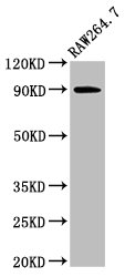
Western Blot Positive WB detected in: Raw264.7 whole cell lysate All lanes: VCP antibody at 3ug/ml Secondary Goat polyclonal to rabbit IgG at 1/50000 dilution Predicted band size: 90 kDa Observed band size: 90 kDa

Western Blot Positive WB detected in: Raw264.7 whole cell lysate All lanes: VCP antibody at 3ug/ml Secondary Goat polyclonal to rabbit IgG at 1/50000 dilution Predicted band size: 90 kDa Observed band size: 90 kDa
VCP Antibody
CSB-PA025813LA01HU
ApplicationsWestern Blot, ELISA, ImmunoHistoChemistry
Product group Antibodies
ReactivityHuman
TargetVCP
Overview
- SupplierCusabio
- Product NameVCP Antibody
- Delivery Days Customer20
- ApplicationsWestern Blot, ELISA, ImmunoHistoChemistry
- CertificationResearch Use Only
- ClonalityPolyclonal
- ConjugateUnconjugated
- Gene ID7415
- Target nameVCP
- Target descriptionvalosin containing protein
- Target synonymsCDC48, FTDALS6, TERA, p97, transitional endoplasmic reticulum ATPase, 15S Mg(2+)-ATPase p97 subunit, TER ATPase
- HostRabbit
- IsotypeIgG
- Protein IDP55072
- Protein NameTransitional endoplasmic reticulum ATPase
- Scientific DescriptionNecessary for the fragmentation of Golgi stacks during mitosis and for their reassembly after mitosis. Involved in the formation of the transitional endoplasmic reticulum (tER). The transfer of membranes from the endoplasmic reticulum to the Golgi apparatus occurs via 50-70 nm transition vesicles which derive from part-rough, part-smooth transitional elements of the endoplasmic reticulum (tER). Vesicle budding from the tER is an ATP-dependent process. The ternary complex containing UFD1L, VCP and NPLOC4 binds ubiquitinated proteins and is necessary for the export of misfolded proteins from the ER to the cytoplasm, where they are degraded by the proteasome. The NPLOC4-UFD1L-VCP complex regulates spindle disassembly at the end of mitosis and is necessary for the formation of a closed nuclear envelope. Regulates E3 ubiquitin-protein ligase activity of RNF19A. Component of the VCP/p97-AMFR/gp78 complex that participates in the final step of the sterol-mediated ubiquitination and endoplasmic reticulum-associated degradation (ERAD) of HMGCR. Involved in endoplasmic reticulum stress-induced pre-emptive quality control, a mechanism that selectively attenuates the translocation of newly synthesized proteins into the endoplasmic reticulum and reroutes them to the cytosol for proteasomal degradation (PubMed:26565908). Also involved in DNA damage response: recruited to double-strand breaks (DSBs) sites in a RNF8- and RNF168-dependent manner and promotes the recruitment of TP53BP1 at DNA damage sites (PubMed:22020440, PubMed:22120668). Recruited to stalled replication forks by SPRTN: may act by mediating extraction of DNA polymerase eta (POLH) to prevent excessive translesion DNA synthesis and limit the incidence of mutations induced by DNA damage (PubMed:23042607, PubMed:23042605). Required for cytoplasmic retrotranslocation of stressed/damaged mitochondrial outer-membrane proteins and their subsequent proteasomal degradation (PubMed:16186510, PubMed:21118995). Essential for the maturation of ubiquitin-containing autophagosomes and the clearance of ubiquitinated protein by autophagy (PubMed:20104022). Acts as a negative regulator of type I interferon production by interacting with DDX58/RIG-I: interaction takes place when DDX58/RIG-I is ubiquitinated via Lys-63-linked ubiquitin on its CARD domains, leading to recruit RNF125 and promote ubiquitination and degradation of DDX58/RIG-I (PubMed:26471729).
- ReactivityHuman
- Storage Instruction-20°C or -80°C
- UNSPSC41116161
Related products
Product group Antibodies
Anti-VCP AntibodyA98420
ApplicationsWestern Blot, ELISA
ReactivityHuman, Mouse, Rat
- SizePrice

Product group Antibodies
Anti-VCP Antibody Picoband(r)A00610-2-CARRIER-FREE
ApplicationsFlow Cytometry, Western Blot, ELISA, ImmunoHistoChemistry
ReactivityHuman, Monkey, Mouse, Rat
TargetVCP
- SizePrice
Product group Antibodies
Anti-VCP Antibody144-60308
ApplicationsWestern Blot, ImmunoHistoChemistry
ReactivityHuman, Mouse, Rat
TargetVCP
- SizePrice
Product group Antibodies

VCP AntibodyLS-C830390
ApplicationsWestern Blot, ELISA, ImmunoHistoChemistry
ReactivityHuman, Mouse, Rat
TargetVCP
- SizePrice
Product group Antibodies
VCP Recombinant Antibody, AbBy Fluor-350 ConjugatedBSM-61451R-BF350
ApplicationsFlow Cytometry, ImmunoFluorescence, Western Blot
ReactivityHuman, Mouse, Rat
TargetVCP
- SizePrice
Product group Antibodies
References

Goat anti-CDC48 / YDL126CEB11800
ApplicationsWestern Blot, ELISA, ImmunoHistoChemistry
ReactivityBovine, Canine, Human, Mouse, Rat, Yeast
TargetVCP
- SizePrice
Product group Antibodies
VCP Polyclonal AntibodyCAC15070
ApplicationsWestern Blot, ELISA, ImmunoHistoChemistry
TargetVCP
- SizePrice
Product group Antibodies
VCP antibodyGTX101089
ApplicationsImmunoPrecipitation, Western Blot, ImmunoHistoChemistry, ImmunoHistoChemistry Frozen, ImmunoHistoChemistry Paraffin
ReactivityHuman, Mouse, Rat, Zebra Fish
TargetVCP
- SizePrice