

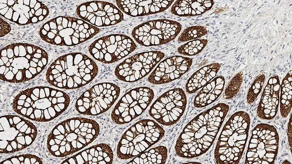
IHC-P analysis of human large bowel tissue using GTX01866 Villin antibody [CWWB1]. Note cytoplasmic staining of the epithelial cells.
IHC-P analysis of human large bowel tissue using GTX01866 Villin antibody [CWWB1]. Note cytoplasmic staining of the epithelial cells.
Villin antibody [CWWB1]
GTX01866
ApplicationsWestern Blot, ImmunoHistoChemistry, ImmunoHistoChemistry Frozen, ImmunoHistoChemistry Paraffin
Product group Antibodies
ReactivityHuman, Porcine, Rat
TargetVIL1
Overview
- SupplierGeneTex
- Product NameVillin antibody [CWWB1]
- Delivery Days Customer9
- Application Supplier NoteWB: 1:500-1:1000. IHC-P: 1:100-1:200. *Optimal dilutions/concentrations should be determined by the researcher.Not tested in other applications.
- ApplicationsWestern Blot, ImmunoHistoChemistry, ImmunoHistoChemistry Frozen, ImmunoHistoChemistry Paraffin
- CertificationResearch Use Only
- ClonalityMonoclonal
- Clone IDCWWB1
- ConjugateUnconjugated
- Gene ID7429
- Target nameVIL1
- Target descriptionvillin 1
- Target synonymsD2S1471, VIL, villin-1
- HostMouse
- IsotypeIgG1
- Protein IDP09327
- Protein NameVillin-1
- Scientific DescriptionThis gene encodes a member of a family of calcium-regulated actin-binding proteins. This protein represents a dominant part of the brush border cytoskeleton which functions in the capping, severing, and bundling of actin filaments. Two mRNAs of 2.7 kb and 3.5 kb have been observed; they result from utilization of alternate poly-adenylation signals present in the terminal exon. [provided by RefSeq, Jul 2008]
- ReactivityHuman, Porcine, Rat
- Storage Instruction2°C to 8°C
- UNSPSC41116161
Datasheet
Related products
Product group Antibodies
Anti-Villin AntibodyA14804
ApplicationsWestern Blot
ReactivityHuman, Mouse, Rat
- SizePrice
Product group Antibodies

VIL1 / Villin AntibodyLS-C783283
ApplicationsWestern Blot
ReactivityHuman
TargetVIL1
- SizePrice

Product group Antibodies
villin Recombinant AntibodyBSM-54212R
ApplicationsFlow Cytometry, ImmunoFluorescence, Western Blot, ImmunoCytoChemistry, ImmunoHistoChemistry, ImmunoHistoChemistry Paraffin
ReactivityHuman, Mouse, Rat
TargetVIL1
- SizePrice
Product group Antibodies
Villin (VIL) Polyclonal AntibodyCAU24074
ApplicationsWestern Blot, ImmunoHistoChemistry
ReactivityMouse, Porcine, Rat
TargetVIL1
- SizePrice
Product group Antibodies
VIL1 AntibodyCSB-PA025855ESR1HU
ApplicationsELISA, ImmunoHistoChemistry
ReactivityHuman
TargetVIL1
- SizePrice
Product group Antibodies
Villin antibodyGTX133791
ApplicationsImmunoFluorescence, Western Blot, ImmunoCytoChemistry
ReactivityHuman, Mouse
TargetVIL1
- SizePrice
![IHC-P analysis of human small intestinal carcinoma section using GTX02738 Villin antibody [rVIL1/1325].](https://www.genetex.com/upload/website/prouct_img/normal/GTX02738/GTX02738_20210319_IHC-P_w_23053122_803.webp)
Product group Antibodies
Villin antibody [rVIL1/1325]GTX02738
ApplicationsImmunoHistoChemistry, ImmunoHistoChemistry Paraffin, Other Application
ReactivityHuman
TargetVIL1
- SizePrice
![IHC-P analysis of human colon adenocarcinoma section using GTX02739 Villin antibody [VIL1/4107R].](https://www.genetex.com/upload/website/prouct_img/normal/GTX02739/GTX02739_20210319_IHC-P_1_w_23053122_200.webp)
Product group Antibodies
Villin antibody [VIL1/4107R]GTX02739
ApplicationsImmunoHistoChemistry, ImmunoHistoChemistry Paraffin
ReactivityHuman
TargetVIL1
- SizePrice
![IHC-P analysis of human small intestine tissue section using GTX02740 Villin antibody [VIL1/2310R].](https://www.genetex.com/upload/website/prouct_img/normal/GTX02740/GTX02740_20210319_IHC-P_w_23053122_374.webp)
Product group Antibodies
Villin antibody [VIL1/2310R]GTX02740
ApplicationsImmunoHistoChemistry, ImmunoHistoChemistry Paraffin
ReactivityHuman
TargetVIL1
- SizePrice
![IHC-P analysis of human renal cortex tissue using GTX04400 Villin antibody [MSVA-459R] HistoMAX?. A strong villin positivity is seen in proximal tubuli of the kidney but not in distal tubuli or glomeruli.](https://www.genetex.com/upload/website/prouct_img/normal/GTX04400/GTX04400_20230728_IHC-P_126_23072722_399.webp)
Product group Antibodies
ApplicationsImmunoHistoChemistry, ImmunoHistoChemistry Paraffin
ReactivityHuman
TargetVIL1
- SizePrice