

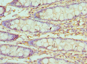
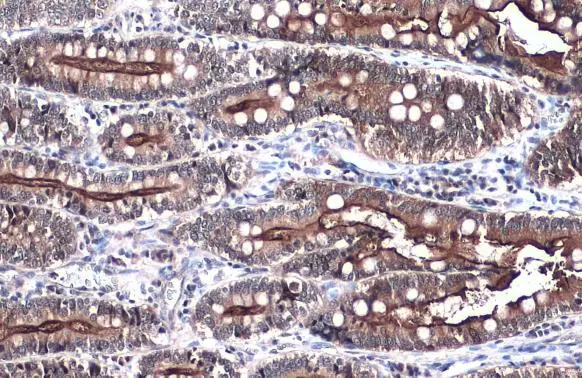
Immunohistochemistry of paraffin-embedded human colon cancer using CSB-PA025855ESR1HU at dilution of 1:100
Immunohistochemistry of paraffin-embedded human colon cancer using CSB-PA025855ESR1HU at dilution of 1:100
VIL1 Antibody
CSB-PA025855ESR1HU
ApplicationsELISA, ImmunoHistoChemistry
Product group Antibodies
ReactivityHuman
TargetVIL1
Overview
- SupplierCusabio
- Product NameVIL1 Antibody
- Delivery Days Customer20
- ApplicationsELISA, ImmunoHistoChemistry
- CertificationResearch Use Only
- ClonalityPolyclonal
- ConjugateUnconjugated
- Gene ID7429
- Target nameVIL1
- Target descriptionvillin 1
- Target synonymsD2S1471, VIL, villin-1
- HostRabbit
- IsotypeIgG
- Protein IDP09327
- Protein NameVillin-1
- Scientific DescriptionEpithelial cell-specific Ca2+-regulated actin-modifying protein that modulates the reorganization of microvillar actin filaments. Plays a role in the actin nucleation, actin filament bundle assembly, actin filament capping and severing. Binds phosphatidylinositol 4,5-bisphosphate (PIP2) and lysophosphatidic acid (LPA); binds LPA with higher affinity than PIP2. Binding to LPA increases its phosphorylation by SRC and inhibits all actin-modifying activities. Binding to PIP2 inhibits actin-capping and -severing activities but enhances actin-bundling activity. Regulates the intestinal epithelial cell morphology, cell invasion, cell migration and apoptosis. Protects against apoptosis induced by dextran sodium sulfate (DSS) in the gastrointestinal epithelium. Appears to regulate cell death by maintaining mitochondrial integrity. Enhances hepatocyte growth factor (HGF)-induced epithelial cell motility, chemotaxis and wound repair. Upon S.flexneri cell infection, its actin-severing activity enhances actin-based motility of the bacteria and plays a role during the dissemination.
- ReactivityHuman
- Storage Instruction-20°C or -80°C
- UNSPSC41116161
Related products
Product group Antibodies
Anti-Villin AntibodyA14804
ApplicationsWestern Blot
ReactivityHuman, Mouse, Rat
- SizePrice
Product group Antibodies

VIL1 / Villin AntibodyLS-C783283
ApplicationsWestern Blot
ReactivityHuman
TargetVIL1
- SizePrice

Product group Antibodies
villin Recombinant AntibodyBSM-54212R
ApplicationsFlow Cytometry, ImmunoFluorescence, Western Blot, ImmunoCytoChemistry, ImmunoHistoChemistry, ImmunoHistoChemistry Paraffin
ReactivityHuman, Mouse, Rat
TargetVIL1
- SizePrice
Product group Antibodies
Villin (VIL) Polyclonal AntibodyCAU24074
ApplicationsWestern Blot, ImmunoHistoChemistry
ReactivityMouse, Porcine, Rat
TargetVIL1
- SizePrice

Product group Antibodies
Anti-VIL1 AntibodyHPA006884
ApplicationsWestern Blot, ImmunoCytoChemistry, ImmunoHistoChemistry
ReactivityHuman
TargetVIL1
- SizePrice
Product group Antibodies
Villin antibodyGTX109940
ApplicationsImmunoFluorescence, Western Blot, ImmunoCytoChemistry, ImmunoHistoChemistry, ImmunoHistoChemistry Frozen, ImmunoHistoChemistry Paraffin
ReactivityCanine, Feline, Human, Mouse, Rat
TargetVIL1
- SizePrice

Product group Antibodies
Anti-Villin/VIL1 Antibody Picoband(r)PB9457-CARRIER-FREE
ApplicationsFlow Cytometry, ImmunoFluorescence, Western Blot, ImmunoHistoChemistry
ReactivityHamster, Human, Mouse, Rat
TargetVIL1
- SizePrice
Product group Antibodies
Anti-Villin1 AntibodyCAB5494
ApplicationsWestern Blot, ELISA
ReactivityHuman
TargetVIL1
- SizePrice