

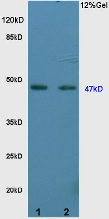
L1 rat brain lysates L2 rat kidney lysates probed with Anti Vitamin D Receptor/VDR Polyclonal Antibody, Unconjugated (bs-2987R) at 1:200 overnight at 4˚C. Followed by conjugation to secondary antibody (bs-0295G-HRP) at 1:3000 for 90 min at 37˚C. Predicted band 47kD. Observed band size:47kD.\n
 for 15min; Block endogenous peroxidase by 3% hydrogen peroxide for 20 minutes; Blocking buffer (normal goat serum) at 37°C for 30min; Antibody incubation with Vitamin D Receptor/VDR Polyclonal Antibody, Unconjugated (bs-2987R) at 1:400 overnight at 4°C, DAB staining.")
at 1:100 dilution in blocking buffer and incubated for 30 min at room temperature, washed twice with 2%BSA in PBS, followed by secondary antibody incubation for 40 min at room temperature. Acquisitions of 20,000 events were performed. Cells stained with primary antibody (green), and isotype control (orange).")
 at 37°C for 20 min; Antibody incubation with (Vitamin D Receptor) polyclonal Antibody, Unconjugated (bs-2987R) 1:100, 90 minutes at 37°C; followed by a conjugated Goat Anti-Rabbit IgG antibody at 37°C for 90 minutes, DAPI (blue, C02-04002) was used to stain the cell nuclei.")
 at 1:200 followed by conjugation to the secondary antibody and DAB staining.")
L1 rat brain lysates L2 rat kidney lysates probed with Anti Vitamin D Receptor/VDR Polyclonal Antibody, Unconjugated (bs-2987R) at 1:200 overnight at 4˚C. Followed by conjugation to secondary antibody (bs-0295G-HRP) at 1:3000 for 90 min at 37˚C. Predicted band 47kD. Observed band size:47kD.\n
Vitamin D Receptor Polyclonal Antibody
BS-2987R
ApplicationsImmunoFluorescence, Western Blot, ImmunoCytoChemistry, ImmunoHistoChemistry, ImmunoHistoChemistry Frozen, ImmunoHistoChemistry Paraffin
Product group Antibodies
ReactivityBovine, Chicken, Equine, Human, Mouse, Porcine, Rabbit, Rat
TargetVDR
Overview
- SupplierBioss
- Product NameVitamin D Receptor Polyclonal Antibody
- Delivery Days Customer16
- ApplicationsImmunoFluorescence, Western Blot, ImmunoCytoChemistry, ImmunoHistoChemistry, ImmunoHistoChemistry Frozen, ImmunoHistoChemistry Paraffin
- Applications SupplierWB(1:300-5000), IHC-P(1:200-400), IHC-F(1:100-500), IF(IHC-P)(1:50-200), IF(IHC-F)(1:50-200), IF(ICC)(1:50-200)
- CertificationResearch Use Only
- ClonalityPolyclonal
- Concentration1 ug/ul
- ConjugateUnconjugated
- Gene ID7421
- Target nameVDR
- Target descriptionvitamin D receptor
- Target synonymsNR1I1, PPP1R163, vitamin D3 receptor, 1,25-dihydroxyvitamin D3 receptor, nuclear receptor subfamily 1 group I member 1, protein phosphatase 1, regulatory subunit 163, vitamin D (1,25- dihydroxyvitamin D3) receptor
- HostRabbit
- IsotypeIgG
- Protein IDP11473
- Protein NameVitamin D3 receptor
- ReactivityBovine, Chicken, Equine, Human, Mouse, Porcine, Rabbit, Rat
- Storage Instruction-20°C
- UNSPSC41116161
References
- Onset of calciotropic receptors during the initiation of mandibular/alveolar bone formation. Bobek J et al., 2020 Jan, Ann AnatRead this paper
- Vitamin D3 mediated regulation of steroidogenesis mitigates testicular activity in an aged rat model. Jeremy M et al., 2019 Jun, J Steroid Biochem Mol BiolRead this paper
- Serum 25-hydroxyvitamin D inversely associated with blood eosinophils in patients with persistent allergic rhinitis. Wu HY et al., 2017 Oct, Asia Pac AllergyRead this paper
- 1,25-Dihydroxyvitamin D3 Promotes High Glucose-Induced M1 Macrophage Switching to M2 via the VDR-PPARgamma Signaling Pathway. Zhang X et al., 2015, Biomed Res IntRead this paper
Datasheet
Related products
Product group Antibodies
Anti-VDR AntibodyA40124
ApplicationsImmunoFluorescence, Western Blot
ReactivityHuman, Mouse, Rat
- SizePrice
Product group Antibodies

ApplicationsWestern Blot, ELISA
- SizePrice
Product group Antibodies
Anti-VDR Antibody144-02194
ApplicationsImmunoFluorescence, Western Blot, ImmunoHistoChemistry
ReactivityHuman, Mouse, Rat
TargetVDR
- SizePrice

Product group Antibodies
Anti-Vitamin D Receptor/VDR Antibody Picoband(r)A00210-1-CARRIER-FREE
ApplicationsFlow Cytometry, Western Blot
ReactivityHuman, Mouse, Rat
TargetVDR
- SizePrice
Product group Antibodies
VDR AntibodyCSB-PA004443
ApplicationsImmunoFluorescence, Western Blot, ELISA, ImmunoHistoChemistry
ReactivityHuman
TargetVDR
- SizePrice
Product group Antibodies

Goat anti-VDREB06803
ApplicationsFlow Cytometry, Western Blot, ELISA
ReactivityCanine, Human, Mouse, Rat
TargetVDR
- SizePrice
Product group Antibodies
ApplicationsImmunoPrecipitation, Western Blot, ImmunoCytoChemistry, ImmunoHistoChemistry
ReactivityMouse
TargetVDR
- SizePrice

Product group Antibodies
Anti-VDR AntibodyHPA047740
ApplicationsImmunoCytoChemistry
ReactivityHuman
TargetVDR
- SizePrice
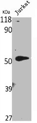
![Various whole cell extracts (30 μg) were separated by 10% SDS-PAGE, and the membrane was blotted with Vitamin D Receptor antibody [HL2661] (GTX639325) diluted at 1:1000. The HRP-conjugated anti-rabbit IgG antibody (GTX213110-01) was used to detect the primary antibody. Corresponding RNA expression data for the same cell lines are based on Human Protein Atlas program.](https://www.genetex.com/upload/website/prouct_img/normal/GTX639325/GTX639325_45271_20231229_WB_TPM_watermark_24010223_174.webp)
Product group Antibodies
Vitamin D Receptor antibody [HL2661]GTX639325
ApplicationsWestern Blot
ReactivityHuman
TargetVDR
- SizePrice