

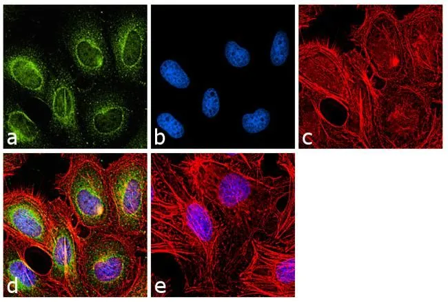
ICC/IF analysis of HeLa cells using GTX79470 VRK1 antibody [1F6]. Panel e is a no primary antibody control. Green : Primary antibody Blue : Nuclei Red : Actin Fixation : 4% paraformaldehyde Permeabilization : 0.1% Triton X-100 for 10 minute Dilution : 1:100 dilution in 0.1% BSA and incubated for 3 hours at room temperature
![ICC/IF analysis of U251 cells using GTX79470 VRK1 antibody [1F6]. Cells were probed without (left) or with(right) an antibody. Green : Primary antibody Blue : Nuclei Red : Actin Fixation : formaldehyde Dilution : 1:200 overnight at 4oC](https://www.genetex.com/upload/website/prouct_img/normal/GTX79470/GTX79470_880_ICC-IF_w_23061322_345.webp "ICC/IF analysis of U251 cells using GTX79470 VRK1 antibody [1F6]. Cells were probed without (left) or with(right) an antibody. Green : Primary antibody Blue : Nuclei Red : Actin Fixation : formaldehyde Dilution : 1:200 overnight at 4oC")
![ICC/IF analysis of murine cells using GTX79470 VRK1 antibody [1F6]. Green : Primary antibody Blue : Nuclei Red : Actin Fixation : formaldehyde Dilution : 1:200 overnight at 4oC](https://www.genetex.com/upload/website/prouct_img/normal/GTX79470/GTX79470_2032_ICC-IF_w_23061322_479.webp "ICC/IF analysis of murine cells using GTX79470 VRK1 antibody [1F6]. Green : Primary antibody Blue : Nuclei Red : Actin Fixation : formaldehyde Dilution : 1:200 overnight at 4oC")
![ICC/IF analysis of HeLa cells using GTX79470 VRK1 antibody [1F6]. Cells were probed without (left) or with(right) an antibody. Green : Primary antibody Blue : Nuclei Red : Actin Fixation : formaldehyde Dilution : 1:200 overnight at 4oC](https://www.genetex.com/upload/website/prouct_img/normal/GTX79470/GTX79470_881_ICC-IF_w_23061322_708.webp "ICC/IF analysis of HeLa cells using GTX79470 VRK1 antibody [1F6]. Cells were probed without (left) or with(right) an antibody. Green : Primary antibody Blue : Nuclei Red : Actin Fixation : formaldehyde Dilution : 1:200 overnight at 4oC")
![ICC/IF analysis of HeLa cells using GTX79470 VRK1 antibody [1F6]. Fixation : Formalin Permeabilization : 0.1% Triton X-100 in TBS for 10 minutes Dilution : 1:100 for at least 1 hour at room temperature](https://www.genetex.com/upload/website/prouct_img/normal/GTX79470/GTX79470_882_ICC-IF_w_23061322_411.webp "ICC/IF analysis of HeLa cells using GTX79470 VRK1 antibody [1F6]. Fixation : Formalin Permeabilization : 0.1% Triton X-100 in TBS for 10 minutes Dilution : 1:100 for at least 1 hour at room temperature")
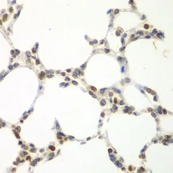
![IHC-P analysis of human testis tissue using GTX79470 VRK1 antibody [1F6]. Right : Primary antibody Left : Negative control without primary antibody Antigen retrieval : heat induced antigen retrieval was performed using 10mM sodium citrate (pH 6.0) buffer microwaved for 8-15 minutes Dilution : 1:200](https://www.genetex.com/upload/website/prouct_img/normal/GTX79470/GTX79470_1400_IHC-P_w_23061322_632.webp "IHC-P analysis of human testis tissue using GTX79470 VRK1 antibody [1F6]. Right : Primary antibody Left : Negative control without primary antibody Antigen retrieval : heat induced antigen retrieval was performed using 10mM sodium citrate (pH 6.0) buffer microwaved for 8-15 minutes Dilution : 1:200")
![IHC-P analysis of human liver tissue using GTX79470 VRK1 antibody [1F6]. Right : Primary antibody Left : Negative control without primary antibody Antigen retrieval : heat induced antigen retrieval was performed using 10mM sodium citrate (pH 6.0) buffer microwaved for 8-15 minutes Dilution : 1:200](https://www.genetex.com/upload/website/prouct_img/normal/GTX79470/GTX79470_1399_IHC-P_w_23061322_964.webp "IHC-P analysis of human liver tissue using GTX79470 VRK1 antibody [1F6]. Right : Primary antibody Left : Negative control without primary antibody Antigen retrieval : heat induced antigen retrieval was performed using 10mM sodium citrate (pH 6.0) buffer microwaved for 8-15 minutes Dilution : 1:200")
![IHC-P analysis of human tonsil tissue using GTX79470 VRK1 antibody [1F6]. Right : Primary antibody Left : Negative control without primary antibody Antigen retrieval : heat induced antigen retrieval was performed using 10mM sodium citrate (pH 6.0) buffer microwaved for 8-15 minutes Dilution : 1:200](https://www.genetex.com/upload/website/prouct_img/normal/GTX79470/GTX79470_1401_IHC-P_w_23061322_581.webp "IHC-P analysis of human tonsil tissue using GTX79470 VRK1 antibody [1F6]. Right : Primary antibody Left : Negative control without primary antibody Antigen retrieval : heat induced antigen retrieval was performed using 10mM sodium citrate (pH 6.0) buffer microwaved for 8-15 minutes Dilution : 1:200")
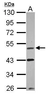
![WB analysis of 25 μg of the indicated whole cell lysates using GTX79470 VRK1 antibody [1F6]. Dilution : 1:750](https://www.genetex.com/upload/website/prouct_img/normal/GTX79470/GTX79470_1942_WB_w_23061322_774.webp "WB analysis of 25 μg of the indicated whole cell lysates using GTX79470 VRK1 antibody [1F6]. Dilution : 1:750")
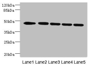
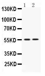
![WB analysis of whole cell extracts (30 ug lysate) of HeLa (Lane 1), HEK 293 (Lane 2), MCF7 (Lane 3), and Jurkat (Lane 4) using GTX79470 VRK1 antibody [1F6]. Dilution : 1:500](https://www.genetex.com/upload/website/prouct_img/normal/GTX79470/GTX79470_1943_WB_w_23061322_789.webp "WB analysis of whole cell extracts (30 ug lysate) of HeLa (Lane 1), HEK 293 (Lane 2), MCF7 (Lane 3), and Jurkat (Lane 4) using GTX79470 VRK1 antibody [1F6]. Dilution : 1:500")
ICC/IF analysis of HeLa cells using GTX79470 VRK1 antibody [1F6]. Panel e is a no primary antibody control. Green : Primary antibody Blue : Nuclei Red : Actin Fixation : 4% paraformaldehyde Permeabilization : 0.1% Triton X-100 for 10 minute Dilution : 1:100 dilution in 0.1% BSA and incubated for 3 hours at room temperature
VRK1 antibody [1F6]
GTX79470
ApplicationsImmunoFluorescence, Western Blot, ImmunoCytoChemistry, ImmunoHistoChemistry, ImmunoHistoChemistry Paraffin
Product group Antibodies
ReactivityHuman, Mouse
TargetVRK1
Overview
- SupplierGeneTex
- Product NameVRK1 antibody [1F6]
- Delivery Days Customer9
- Application Supplier NoteWB: 1:500. ICC/IF: 1:100. IHC-P: 1:100 - 1:500. *Optimal dilutions/concentrations should be determined by the researcher.Not tested in other applications.
- ApplicationsImmunoFluorescence, Western Blot, ImmunoCytoChemistry, ImmunoHistoChemistry, ImmunoHistoChemistry Paraffin
- CertificationResearch Use Only
- ClonalityMonoclonal
- Clone ID1F6
- ConjugateUnconjugated
- Gene ID7443
- Target nameVRK1
- Target descriptionVRK serine/threonine kinase 1
- Target synonymsHMNR10, PCH1, PCH1A, serine/threonine-protein kinase VRK1, vaccinia related kinase 1, vaccinia virus B1R-related kinase 1
- HostMouse
- IsotypeIgG1
- Protein IDQ99986
- Protein NameSerine/threonine-protein kinase VRK1
- Scientific DescriptionThis gene encodes a member of the vaccinia-related kinase (VRK) family of serine/threonine protein kinases. This gene is widely expressed in human tissues and has increased expression in actively dividing cells, such as those in testis, thymus, fetal liver, and carcinomas. Its protein localizes to the nucleus and has been shown to promote the stability and nuclear accumulation of a transcriptionally active p53 molecule and, in vitro, to phosphorylate Thr18 of p53 and reduce p53 ubiquitination. This gene, therefore, may regulate cell proliferation. This protein also phosphorylates histone, casein, and the transcription factors ATF2 (activating transcription factor 2) and c-JUN. [provided by RefSeq, Jul 2008]
- ReactivityHuman, Mouse
- Storage Instruction2°C to 8°C
- UNSPSC41116161
Datasheet
Related products
Product group Antibodies
Anti-VRK1 Antibody144-07745
ApplicationsWestern Blot, ImmunoHistoChemistry
ReactivityHuman, Mouse, Rat
TargetVRK1
- SizePrice

Product group Antibodies
Anti-VRK1 AntibodyA44505
ApplicationsWestern Blot, ImmunoHistoChemistry
ReactivityHuman
- SizePrice
Product group Antibodies

VRK1 AntibodyLS-C831877
ApplicationsWestern Blot, ELISA, ImmunoHistoChemistry
ReactivityHuman, Mouse
TargetVRK1
- SizePrice

Product group Antibodies
Anti-VRK1 AntibodyHPA000660
ApplicationsWestern Blot
ReactivityHuman
TargetVRK1
- SizePrice
Product group Antibodies
VRK1 AntibodyCSB-PA857466DSR1HU
ApplicationsWestern Blot, ELISA, ImmunoHistoChemistry
ReactivityHuman
TargetVRK1
- SizePrice
Product group Antibodies
VRK1 Polyclonal AntibodyBS-7706R
ApplicationsImmunoFluorescence, Western Blot, ELISA, ImmunoCytoChemistry, ImmunoHistoChemistry, ImmunoHistoChemistry Frozen, ImmunoHistoChemistry Paraffin
ReactivityBovine, Equine, Human, Mouse, Porcine, Rat, Sheep
TargetVRK1
- SizePrice
Product group Antibodies
VRK1 antibodyGTX107899
ApplicationsWestern Blot, ImmunoHistoChemistry, ImmunoHistoChemistry Paraffin
ReactivityHuman
TargetVRK1
- SizePrice
Product group Antibodies
VRK1 antibodyGTX33581
ApplicationsWestern Blot, ImmunoHistoChemistry, ImmunoHistoChemistry Paraffin
ReactivityHuman, Mouse, Rat
TargetVRK1
- SizePrice
Product group Antibodies
Anti-VRK1 Antibody Picoband(r)PB9907-CARRIER-FREE
ApplicationsWestern Blot
ReactivityHuman, Rat
TargetVRK1
- SizePrice