

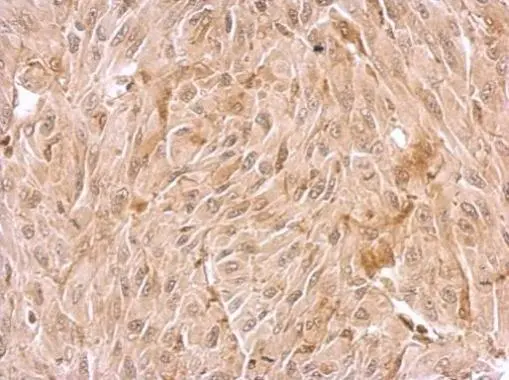
WDR1 antibody
70R-3385
Product group Antibodies
Overview
- SupplierBiosynth
- Product NameWDR1 antibody
- Delivery Days Customer7
- CertificationResearch Use Only
- UNSPSC41116161
Related products
Product group Antibodies
Anti-WDR1 Antibody144-12163
ApplicationsWestern Blot
ReactivityHuman, Mouse, Rat
TargetWDR1
- SizePrice
Product group Antibodies

WDR1 AntibodyLS-C832556
ApplicationsWestern Blot, ELISA
ReactivityHuman, Mouse, Rat
TargetWDR1
- SizePrice
Product group Antibodies
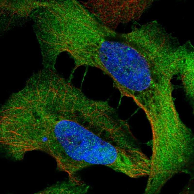
WDR1 Recombinant AntibodyBSM-62672R
ApplicationsImmunoFluorescence, Western Blot, ImmunoCytoChemistry, ImmunoHistoChemistry, ImmunoHistoChemistry Frozen, ImmunoHistoChemistry Paraffin
ReactivityHuman
TargetWDR1
- SizePrice
Product group Antibodies
WDR1 AntibodyCSB-PA025995GA01HU
ApplicationsWestern Blot, ELISA, ImmunoHistoChemistry
ReactivityHuman, Mouse, Rat
TargetWDR1
- SizePrice
Product group Antibodies
WDR1 antibodyGTX102214
ApplicationsWestern Blot, ImmunoHistoChemistry, ImmunoHistoChemistry Paraffin
ReactivityHuman, Mouse
TargetWDR1
- SizePrice
Product group Antibodies
Anti-WDR1 AntibodyHPA070293
ApplicationsWestern Blot, ImmunoCytoChemistry
ReactivityHuman
TargetWDR1
- SizePrice

Product group Antibodies
Anti-WDR1 Antibody Picoband(r)PB9962-CARRIER-FREE
ApplicationsWestern Blot
ReactivityBovine, Human, Mouse, Rat
TargetWDR1
- SizePrice