

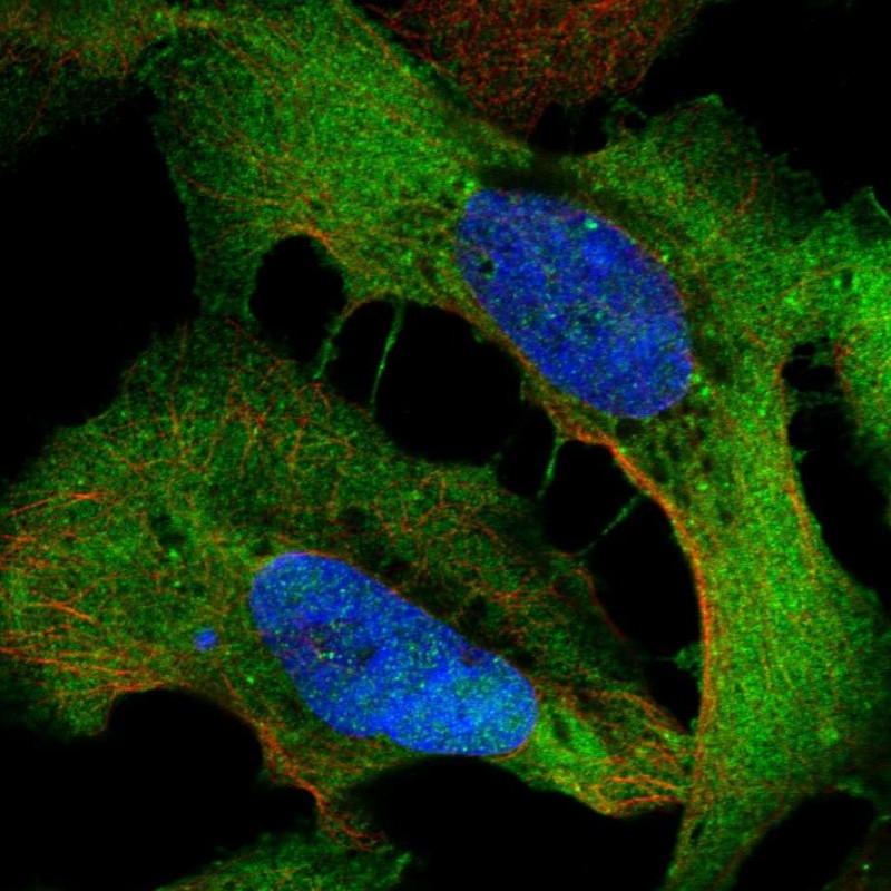
Immunofluorescent staining of human cell line U-2 OS shows localization to plasma membrane & cell junctions.

Immunofluorescent staining of human cell line U-2 OS shows localization to plasma membrane & cell junctions.
Anti-WDR1 Antibody
HPA070293
ApplicationsWestern Blot, ImmunoCytoChemistry
Product group Antibodies
ReactivityHuman
TargetWDR1
Overview
- SupplierAtlas Antibodies
- Product NameAnti-WDR1 Antibody
- Delivery Days Customer4
- ApplicationsWestern Blot, ImmunoCytoChemistry
- CertificationResearch Use Only
- ClonalityPolyclonal
- ConjugateUnconjugated
- Gene ID9948
- Target nameWDR1
- Target descriptionWD repeat domain 1
- Target synonymsAIP1, HEL-S-52, NORI-1, PFITS, WD repeat-containing protein 1, actin-interacting protein 1, epididymis secretory protein Li 52
- HostRabbit
- IsotypeIgG
- Protein IDO75083
- Protein NameWD repeat-containing protein 1
- Scientific DescriptionRecombinant Protein Epitope Signature Tag (PrEST) antigen sequence
- ReactivityHuman
- Storage Instruction-20°C,2°C to 8°C
- UNSPSC41116161
Datasheet
MSDS
Related products
Product group Antibodies
Anti-WDR1 Antibody144-12163
ApplicationsWestern Blot
ReactivityHuman, Mouse, Rat
TargetWDR1
- SizePrice
Product group Antibodies

WDR1 AntibodyLS-C832556
ApplicationsWestern Blot, ELISA
ReactivityHuman, Mouse, Rat
TargetWDR1
- SizePrice
Product group Antibodies
WDR1 Recombinant AntibodyBSM-62672R
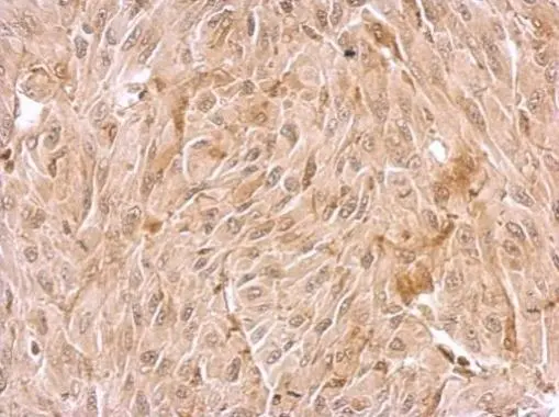
ApplicationsImmunoFluorescence, Western Blot, ImmunoCytoChemistry, ImmunoHistoChemistry, ImmunoHistoChemistry Frozen, ImmunoHistoChemistry Paraffin
ReactivityHuman
TargetWDR1
- SizePrice
Product group Antibodies
WDR1 AntibodyCSB-PA025995GA01HU
ApplicationsWestern Blot, ELISA, ImmunoHistoChemistry
ReactivityHuman, Mouse, Rat
TargetWDR1
- SizePrice
Product group Antibodies
WDR1 antibodyGTX102214
ApplicationsWestern Blot, ImmunoHistoChemistry, ImmunoHistoChemistry Paraffin
ReactivityHuman, Mouse
TargetWDR1
- SizePrice

Product group Antibodies
Anti-WDR1 AntibodyHPA036434
ApplicationsWestern Blot, ImmunoHistoChemistry
ReactivityHuman
TargetWDR1
- SizePrice