

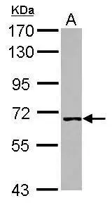
Western blot analysis is shown using GeneTex Affinity Purified anti-Human WHIP antibody (GTX24731) to detect Human WHIP present in a HEK293 whole cell lysate. ~30mg of lysate was loaded per lane for 4-20% gradient SDS-PAGE. Comparison to a molecular weight marker (not shown) indicates a primary band of ~96.0 kDa is detected. The identity of the minor band migrating at a slightly higher molecular weight is unknown, but may represent an alternate isoform of WHIP or post translational modification of the WHIP protein. The blot was incubated with a 1:200 dilution of the antibody at room temperature for 2 h followed by detection using infrared labeled Goat-a-Rabbit IgG [H&L] MX10 diluted 1:5,000 for 45 min. The fluorescence image was captured using the OdysseyR Infrared Imaging System developed by LI-COR.
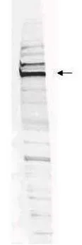
 or without (Lane A) blocking peptide. Loading : 30 μg Dilution : 1:1000")

Western blot analysis is shown using GeneTex Affinity Purified anti-Human WHIP antibody (GTX24731) to detect Human WHIP present in a HEK293 whole cell lysate. ~30mg of lysate was loaded per lane for 4-20% gradient SDS-PAGE. Comparison to a molecular weight marker (not shown) indicates a primary band of ~96.0 kDa is detected. The identity of the minor band migrating at a slightly higher molecular weight is unknown, but may represent an alternate isoform of WHIP or post translational modification of the WHIP protein. The blot was incubated with a 1:200 dilution of the antibody at room temperature for 2 h followed by detection using infrared labeled Goat-a-Rabbit IgG [H&L] MX10 diluted 1:5,000 for 45 min. The fluorescence image was captured using the OdysseyR Infrared Imaging System developed by LI-COR.
WHIP antibody
GTX24731
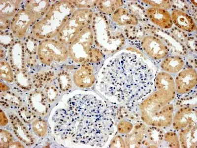
ApplicationsWestern Blot, ELISA, ImmunoHistoChemistry
Product group Antibodies
ReactivityHuman
TargetWRNIP1
Overview
- SupplierGeneTex
- Product NameWHIP antibody
- Delivery Days Customer9
- Application Supplier NoteWB: 1:500-1:2000. ELISA: 1:10000-1:40000. *Optimal dilutions/concentrations should be determined by the researcher.Not tested in other applications.
- ApplicationsWestern Blot, ELISA, ImmunoHistoChemistry
- CertificationResearch Use Only
- ClonalityPolyclonal
- Concentration1 mg/ml
- ConjugateUnconjugated
- Gene ID56897
- Target nameWRNIP1
- Target descriptionWRN helicase interacting protein 1
- Target synonymsCFAP93, FAP93, WHIP, bA420G6.2, ATPase WRNIP1, Werner helicase interacting protein 1, putative helicase RUVBL
- HostRabbit
- IsotypeIgG
- Protein IDQ96S55
- Protein NameATPase WRNIP1
- Scientific DescriptionWHIP Werners syndrome is a rare autosomal recessive disorder characterized by premature aging. Werner helicase interacting protein 1 (WHIP) interacts with the N-terminal portion of Werner protein containing the exonuclease domain. This protein shows homology to replication factor C family proteins, and is conserved from E. coli to human. Studies in yeast suggest that this gene may influence the aging process.
- ReactivityHuman
- Storage Instruction-20°C or -80°C,2°C to 8°C
- UNSPSC41116161
References
- Human Wrnip1 is localized in replication factories in a ubiquitin-binding zinc finger-dependent manner. Crosetto N et al., 2008 Dec 12, J Biol ChemRead this paper
Datasheet
Related products
Product group Antibodies
Anti-WRNIP1 (N-term) Antibody102-23472
ApplicationsWestern Blot
TargetWRNIP1
- SizePrice
Product group Antibodies

Goat anti-WHIP / WRNIP1EB05132
ApplicationsWestern Blot, ELISA, ImmunoHistoChemistry
ReactivityBovine, Canine, Human, Mouse, Rat
TargetWRNIP1
- SizePrice
Product group Antibodies
WRNIP1 AntibodyCSB-PA026150GA01HU
ApplicationsWestern Blot, ELISA
ReactivityHuman, Mouse, Rat
TargetWRNIP1
- SizePrice
Product group Antibodies

WRNIP1 / WHIP AntibodyLS-C410704
ApplicationsWestern Blot
ReactivityHuman
TargetWRNIP1
- SizePrice
Product group Antibodies
WHIP antibody, C-termGTX89707
ApplicationsWestern Blot, ImmunoHistoChemistry, ImmunoHistoChemistry Paraffin
ReactivityHuman
TargetWRNIP1
- SizePrice
Product group Antibodies
WHIP antibody [C1C3]GTX107101
ApplicationsWestern Blot
ReactivityHuman
TargetWRNIP1
- SizePrice

Product group Antibodies
Anti-WRNIP1 AntibodyHPA031753
ApplicationsWestern Blot, ImmunoCytoChemistry, ImmunoHistoChemistry
ReactivityHuman
TargetWRNIP1
- SizePrice