

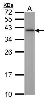
Sample (30 ug of whole cell lysate) A: Hep G2 (GTX27900) 12% SDS PAGE GTX100092 diluted at 1:1000
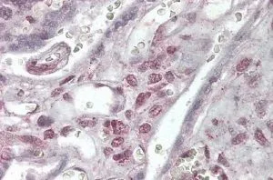
![XLF antibody [N3C3] detects XLF protein on human ovarian carcinoma by immunohistochemical analysis. Sample: Paraffin-embedded ovarian carcinoma. XLF antibody [N3C3] (GTX100092) dilution: 1:500.
Antigen Retrieval: Trilogy? (EDTA based, pH 8.0) buffer, 15min](https://www.genetex.com/upload/website/prouct_img/normal/GTX100092/GTX100092_41031_IHC_w_23053123_248.webp "XLF antibody [N3C3] detects XLF protein on human ovarian carcinoma by immunohistochemical analysis. Sample: Paraffin-embedded ovarian carcinoma. XLF antibody [N3C3] (GTX100092) dilution: 1:500.
Antigen Retrieval: Trilogy? (EDTA based, pH 8.0) buffer, 15min")
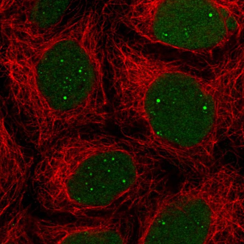
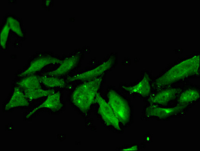
![XLF antibody [N3C3] detects XLF protein at nucleus by immunofluorescent analysis. Sample: HeLa cells were fixed in 4% paraformaldehyde at RT for 15 min. Green: XLF protein stained by XLF antibody [N3C3] (GTX100092) diluted at 1:1000. Blue: Hoechst 33342 staining.](https://www.genetex.com/upload/website/prouct_img/normal/GTX100092/GTX100092_41570_IFA_w_23053123_204.webp "XLF antibody [N3C3] detects XLF protein at nucleus by immunofluorescent analysis. Sample: HeLa cells were fixed in 4% paraformaldehyde at RT for 15 min. Green: XLF protein stained by XLF antibody [N3C3] (GTX100092) diluted at 1:1000. Blue: Hoechst 33342 staining.")
![XLF antibody [N3C3] immunoprecipitates XLF protein in IP experiments. IP samples: Jurkat whole cell extract A. Control with 3 μg of preimmune Rabbit IgG B. Immunoprecipitation of XLF protein by 3 μg XLF antibody [N3C3] (GTX100092) 10 % SDS-PAGE The immunoprecipitated XLF protein was detected by XLF antibody [N3C3] (GTX100092) diluted at 1:1000. [EasyBlot anti-rabbit IgG (GTX221666-01) was used as a secondary reagent]](https://www.genetex.com/upload/website/prouct_img/normal/GTX100092/GTX100092_41570_IP_w_23053123_678.webp "XLF antibody [N3C3] immunoprecipitates XLF protein in IP experiments. IP samples: Jurkat whole cell extract A. Control with 3 μg of preimmune Rabbit IgG B. Immunoprecipitation of XLF protein by 3 μg XLF antibody [N3C3] (GTX100092) 10 % SDS-PAGE The immunoprecipitated XLF protein was detected by XLF antibody [N3C3] (GTX100092) diluted at 1:1000. [EasyBlot anti-rabbit IgG (GTX221666-01) was used as a secondary reagent]")
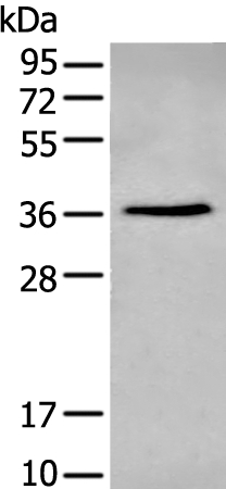
Sample (30 ug of whole cell lysate) A: Hep G2 (GTX27900) 12% SDS PAGE GTX100092 diluted at 1:1000
XLF antibody [N3C3]
GTX100092
ApplicationsImmunoFluorescence, ImmunoPrecipitation, Western Blot, ImmunoCytoChemistry, ImmunoHistoChemistry, ImmunoHistoChemistry Paraffin
Product group Antibodies
ReactivityHuman
TargetNHEJ1
Overview
- SupplierGeneTex
- Product NameXLF antibody [N3C3]
- Delivery Days Customer9
- Application Supplier NoteWB: 1:500-1:3000. ICC/IF: 1:100-1:1000. IHC-P: 1:100-1:1000. IP: 1:100-1:500. *Optimal dilutions/concentrations should be determined by the researcher.Not tested in other applications.
- ApplicationsImmunoFluorescence, ImmunoPrecipitation, Western Blot, ImmunoCytoChemistry, ImmunoHistoChemistry, ImmunoHistoChemistry Paraffin
- CertificationResearch Use Only
- ClonalityPolyclonal
- Concentration1 mg/ml
- ConjugateUnconjugated
- Gene ID79840
- Target nameNHEJ1
- Target descriptionnon-homologous end joining factor 1
- Target synonymsIMD124, MCOPCB13, XLF, non-homologous end-joining factor 1, XRCC4-like factor, nonhomologous end-joining factor 1, protein cernunnos
- HostRabbit
- IsotypeIgG
- Protein IDQ9H9Q4
- Protein NameNon-homologous end-joining factor 1
- Scientific DescriptionDouble-strand breaks in DNA result from genotoxic stresses and are among the most damaging of DNA lesions. This gene encodes a DNA repair factor essential for the nonhomologous end-joining pathway, which preferentially mediates repair of double-stranded breaks. Mutations in this gene cause different kinds of severe combined immunodeficiency disorders. [provided by RefSeq]
- ReactivityHuman
- Storage Instruction-20°C or -80°C,2°C to 8°C
- UNSPSC41116161
Datasheet
Related products

Product group Antibodies
Anti-XLF/NHEJ1 Picoband(r) AntibodyA03552-3-CARRIER-FREE
ApplicationsFlow Cytometry, ImmunoFluorescence, Western Blot, ELISA, ImmunoCytoChemistry, ImmunoHistoChemistry
ReactivityHuman, Mouse
TargetNHEJ1
- SizePrice
Product group Antibodies
Anti-NHEJ1 AntibodyA44469
ApplicationsWestern Blot, ImmunoHistoChemistry
ReactivityHuman
- SizePrice
Product group Antibodies

ApplicationsWestern Blot, ELISA, ImmunoHistoChemistry
ReactivityCanine, Human
TargetNHEJ1
- SizePrice
Product group Antibodies
Anti-NHEJ1 AntibodyHPA043869
ApplicationsImmunoCytoChemistry
ReactivityHuman
TargetNHEJ1
- SizePrice
Product group Antibodies

NHEJ1 / XLF AntibodyLS-C668379
ApplicationsWestern Blot
ReactivityHuman
TargetNHEJ1
- SizePrice
Product group Antibodies
NHEJ1 AntibodyCSB-PA881005LA01HU
ApplicationsImmunoFluorescence, ELISA
ReactivityHuman
TargetNHEJ1
- SizePrice
![WB analysis of HEK293T cells transfected with XLF plasmid (Right) or empty vector (Left) for 48 hrs using GTX84043 XLF antibody [1G7]. Loading : 5 ug per lane](https://www.genetex.com/upload/website/prouct_img/normal/GTX84043/GTX84043_4090_WB_w_23061420_816.webp)
Product group Antibodies
XLF antibody [1G7]GTX84043
ApplicationsFlow Cytometry, ImmunoFluorescence, Western Blot, ImmunoCytoChemistry
ReactivityHuman
TargetNHEJ1
- SizePrice
![WB analysis of HEK293T cells transfected with XLF plasmid (Right) or empty vector (Left) for 48 hrs using GTX84044 XLF antibody [3E5]. Loading : 5 ug per lane](https://www.genetex.com/upload/website/prouct_img/normal/GTX84044/GTX84044_4091_WB_w_23061420_498.webp)
Product group Antibodies
XLF antibody [3E5]GTX84044
ApplicationsFlow Cytometry, Western Blot
ReactivityHuman
TargetNHEJ1
- SizePrice
Product group Antibodies
XLF antibody, C-termGTX89244
ApplicationsWestern Blot, ImmunoHistoChemistry, ImmunoHistoChemistry Paraffin
ReactivityHuman
TargetNHEJ1
- SizePrice
Product group Antibodies
NHEJ1 Recombinant Antibody, AbBy Fluor-555 ConjugatedBSM-62071R-BF555
ApplicationsFlow Cytometry, ImmunoFluorescence, Western Blot
ReactivityHuman, Mouse, Rat
TargetNHEJ1
- SizePrice