

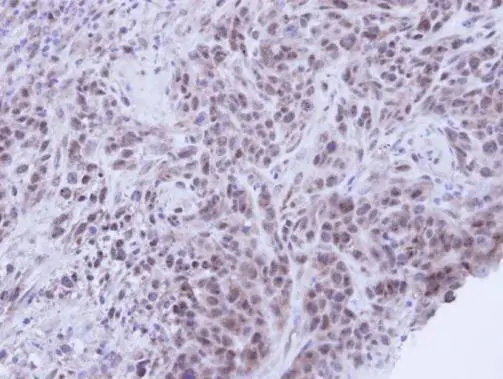
Immunohistochemical analysis of paraffin-embedded SAS Xenograft , using XRCC1(GTX111712) antibody at 1:100 dilution.
Antigen Retrieval: Trilogy? (EDTA based, pH 8.0) buffer, 15min
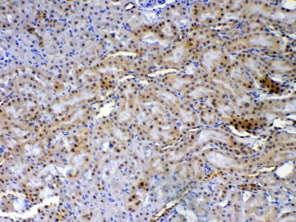
![Rat tissue extract (50 μg) was separated by 7.5% SDS-PAGE, and the membrane was blotted with XRCC1 antibody [N1N3] (GTX111712) diluted at 1:500.](https://www.genetex.com/upload/website/prouct_img/normal/GTX111712/GTX111712_40079_20151119_WB_R_kidney_w_23060500_283.webp "Rat tissue extract (50 μg) was separated by 7.5% SDS-PAGE, and the membrane was blotted with XRCC1 antibody [N1N3] (GTX111712) diluted at 1:500.")
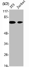
 A: 293T 7.5% SDS PAGE GTX111712 diluted at 1:1000")
 antibody at 1:200 dilution.")
Immunohistochemical analysis of paraffin-embedded SAS Xenograft , using XRCC1(GTX111712) antibody at 1:100 dilution.
Antigen Retrieval: Trilogy? (EDTA based, pH 8.0) buffer, 15min
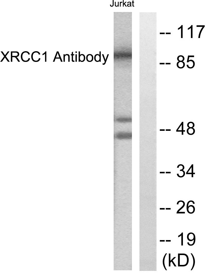
XRCC1 antibody [N1N3]
GTX111712
ApplicationsImmunoFluorescence, Western Blot, ImmunoCytoChemistry, ImmunoHistoChemistry, ImmunoHistoChemistry Paraffin
Product group Antibodies
ReactivityHuman, Rat
TargetXRCC1
Overview
- SupplierGeneTex
- Product NameXRCC1 antibody [N1N3]
- Delivery Days Customer9
- Application Supplier NoteWB: 1:500-1:3000. ICC/IF: 1:100-1:1000. IHC-P: 1:100-1:1000. *Optimal dilutions/concentrations should be determined by the researcher.Not tested in other applications.
- ApplicationsImmunoFluorescence, Western Blot, ImmunoCytoChemistry, ImmunoHistoChemistry, ImmunoHistoChemistry Paraffin
- CertificationResearch Use Only
- ClonalityPolyclonal
- Concentration0.73 mg/ml
- ConjugateUnconjugated
- Gene ID7515
- Target nameXRCC1
- Target descriptionX-ray repair cross complementing 1
- Target synonymsRCC, SCAR26, DNA repair protein XRCC1, X-ray repair complementing defective repair in Chinese hamster cells 1, X-ray repair cross-complementing protein 1
- HostRabbit
- IsotypeIgG
- Protein IDP18887
- Protein NameDNA repair protein XRCC1
- Scientific DescriptionThe protein encoded by this gene is involved in the efficient repair of DNA single-strand breaks formed by exposure to ionizing radiation and alkylating agents. This protein interacts with DNA ligase III, polymerase beta and poly (ADP-ribose) polymerase to participate in the base excision repair pathway. It may play a role in DNA processing during meiogenesis and recombination in germ cells. A rare microsatellite polymorphism in this gene is associated with cancer in patients of varying radiosensitivity. [provided by RefSeq]
- ReactivityHuman, Rat
- Storage Instruction-20°C or -80°C,2°C to 8°C
- UNSPSC41116161
Datasheet
Related products
Product group Antibodies
Anti-XRCC1 AntibodyA95758
ApplicationsWestern Blot, ELISA, ImmunoHistoChemistry
ReactivityHuman, Mouse, Rat
- SizePrice
Product group Antibodies
Anti-XRCC1 Antibody144-61620
ApplicationsImmunoFluorescence, Western Blot
ReactivityHuman, Mouse
TargetXRCC1
- SizePrice
Product group Antibodies
Anti-XRCC1 Antibody Picoband(r)A00571-CARRIER-FREE
ApplicationsFlow Cytometry, ImmunoFluorescence, Western Blot, ELISA, ImmunoCytoChemistry, ImmunoHistoChemistry, ImmunoHistoChemistry Frozen
ReactivityHuman, Mouse, Rat
TargetXRCC1
- SizePrice
Product group Antibodies
XRCC1 Recombinant Antibody, AbBy Fluor-594 ConjugatedBSM-61465R-BF594
ApplicationsWestern Blot
ReactivityHuman, Mouse, Rat
TargetXRCC1
- SizePrice
Product group Antibodies
ApplicationsImmunoPrecipitation, Western Blot, ImmunoCytoChemistry, ImmunoHistoChemistry
TargetXRCC1
- SizePrice
Product group Antibodies
XRCC1 AntibodyCSB-PA004528
ApplicationsWestern Blot, ELISA, ImmunoHistoChemistry
ReactivityHuman, Mouse, Rat
TargetXRCC1
- SizePrice
![IHC-P analysis of human testis tissue using GTX21838 XRCC1 antibody [33-2-5].](https://www.genetex.com/upload/website/prouct_img/normal/GTX21838/GTX21838_20191203_IHC-P_90_w_23060620_921.webp)
Product group Antibodies
References
XRCC1 antibody [33-2-5]GTX21838
ApplicationsImmunoFluorescence, ImmunoCytoChemistry, ImmunoHistoChemistry, ImmunoHistoChemistry Paraffin
ReactivityHuman
TargetXRCC1
- SizePrice
![FACS analysis of Jurkat cells using GTX83410 XRCC1 antibody [2G8]. Red : Primary antibody Blue : Negative control antibody](https://www.genetex.com/upload/website/prouct_img/normal/GTX83410/GTX83410_8_FACS_w_23061419_675.webp)
Product group Antibodies
XRCC1 antibody [2G8]GTX83410
ApplicationsFlow Cytometry, Western Blot
ReactivityHuman
TargetXRCC1
- SizePrice
![IHC-P analysis of human kidney tissue using GTX83411 XRCC1 antibody [2D8]. Antigen retrieval : Heat-induced epitope retrieval by 10mM citrate buffer, pH6.0, 100oC for 10min.](https://www.genetex.com/upload/website/prouct_img/normal/GTX83411/GTX83411_1345_IHC-P_w_23061419_309.webp)
Product group Antibodies
References
XRCC1 antibody [2D8]GTX83411
ApplicationsFlow Cytometry, Western Blot, ImmunoHistoChemistry, ImmunoHistoChemistry Paraffin
ReactivityHuman, Monkey
TargetXRCC1
- SizePrice

Product group Antibodies
Anti-XRCC1 AntibodyHPA006717
ApplicationsWestern Blot, ImmunoCytoChemistry, ImmunoHistoChemistry
ReactivityHuman
TargetXRCC1
- SizePrice