

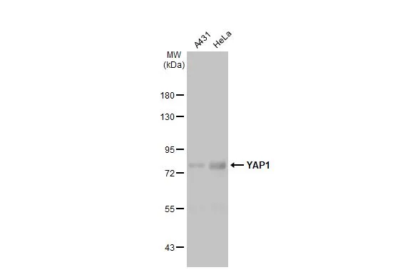
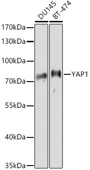
Various whole cell extracts (30 μg) were separated by 7.5% SDS-PAGE, and the membrane was blotted with YAP1 antibody [HL1894] (GTX637643) diluted at 1:4000. The HRP-conjugated anti-rabbit IgG antibody (GTX213110-01) was used to detect the primary antibody.
![Non-transfected (–) and transfected (+) HeLa whole cell extracts (30 μg) were separated by 7.5% SDS-PAGE, and the membrane was blotted with YAP1 antibody [HL1894] (GTX637643) diluted at 1:4000. The HRP-conjugated anti-rabbit IgG antibody (GTX213110-01) was used to detect the primary antibody, and the signal was developed with Trident ECL plus-Enhanced.](https://www.genetex.com/upload/website/prouct_img/normal/GTX637643/GTX637643_T-44837_20221111_WB_shRNA_watermark_22111518_232.webp "Non-transfected (–) and transfected (+) HeLa whole cell extracts (30 μg) were separated by 7.5% SDS-PAGE, and the membrane was blotted with YAP1 antibody [HL1894] (GTX637643) diluted at 1:4000. The HRP-conjugated anti-rabbit IgG antibody (GTX213110-01) was used to detect the primary antibody, and the signal was developed with Trident ECL plus-Enhanced.")
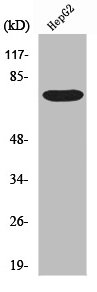
![Various whole cell extracts (30 μg) were separated by 7.5% SDS-PAGE, and the membrane was blotted with YAP1 antibody [HL1894] (GTX637643) diluted at 1:4000. The HRP-conjugated anti-rabbit IgG antibody (GTX213110-01) was used to detect the primary antibody, and the signal was developed with Trident ECL plus-Enhanced. Corresponding RNA expression data for the same cell lines are based on Human Protein Atlas program.](https://www.genetex.com/upload/website/prouct_img/normal/GTX637643/GTX637643_T-44837_20221125_WB_TPM_watermark_22112219_365.webp "Various whole cell extracts (30 μg) were separated by 7.5% SDS-PAGE, and the membrane was blotted with YAP1 antibody [HL1894] (GTX637643) diluted at 1:4000. The HRP-conjugated anti-rabbit IgG antibody (GTX213110-01) was used to detect the primary antibody, and the signal was developed with Trident ECL plus-Enhanced. Corresponding RNA expression data for the same cell lines are based on Human Protein Atlas program.")
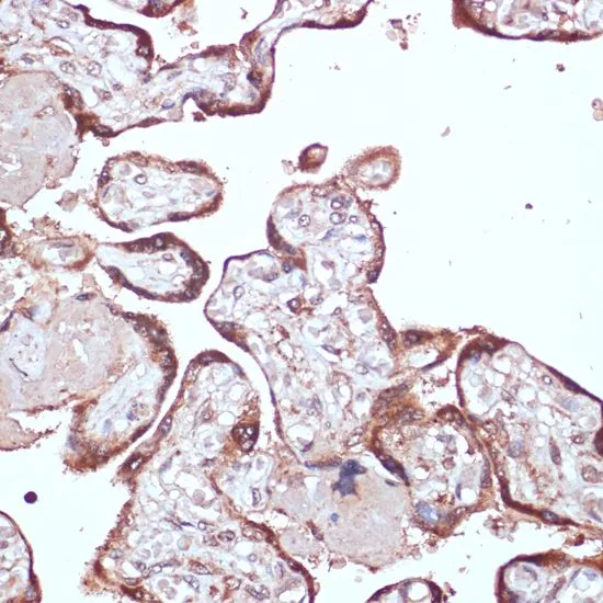
![YAP1 antibody [HL1894] detects YAP1 protein at cytoplasm by immunohistochemical analysis. Sample: Paraffin-embedded human colon cancer. YAP1 stained by YAP1 antibody [HL1894] (GTX637643) diluted at 1:100. Antigen Retrieval: Citrate buffer, pH 6.0, 15 min](https://www.genetex.com/upload/website/prouct_img/normal/GTX637643/GTX637643_T-44837_20221215_IHC-P_22122722_770.webp "YAP1 antibody [HL1894] detects YAP1 protein at cytoplasm by immunohistochemical analysis. Sample: Paraffin-embedded human colon cancer. YAP1 stained by YAP1 antibody [HL1894] (GTX637643) diluted at 1:100. Antigen Retrieval: Citrate buffer, pH 6.0, 15 min")
![YAP1 antibody [HL1894] detects YAP1 protein by immunofluorescent analysis. Sample: U2OS cells were fixed in 4% paraformaldehyde at RT for 15 min. Green: YAP1 stained by YAP1 antibody [HL1894] (GTX637643) diluted at 1:500. Red: alpha Tubulin, a cytoskeleton marker, stained by alpha Tubulin antibody [GT114] (GTX628802) diluted at 1:1000.](https://www.genetex.com/upload/website/prouct_img/normal/GTX637643/GTX637643_44900_20221230_ICC_IF_23010219_424.webp "YAP1 antibody [HL1894] detects YAP1 protein by immunofluorescent analysis. Sample: U2OS cells were fixed in 4% paraformaldehyde at RT for 15 min. Green: YAP1 stained by YAP1 antibody [HL1894] (GTX637643) diluted at 1:500. Red: alpha Tubulin, a cytoskeleton marker, stained by alpha Tubulin antibody [GT114] (GTX628802) diluted at 1:1000.")
![Various whole cell extracts (30 μg) were separated by 7.5% SDS-PAGE, and the membranes were blotted with YAP1 antibody [HL1894] (GTX637643) diluted at 1:1000 and competitor's antibody (#Highly competitor antibody) diluted at 1:1000. The HRP-conjugated anti-rabbit IgG antibody (GTX213110-01) was used to detect the primary antibody. *The competitor is not affiliated with GeneTex and does not endorse this product.](https://www.genetex.com/upload/website/prouct_img/normal/GTX637643/GTX637643_44900_20230707_WB_competitor_watermark_23071223_996.webp "Various whole cell extracts (30 μg) were separated by 7.5% SDS-PAGE, and the membranes were blotted with YAP1 antibody [HL1894] (GTX637643) diluted at 1:1000 and competitor's antibody (#Highly competitor antibody) diluted at 1:1000. The HRP-conjugated anti-rabbit IgG antibody (GTX213110-01) was used to detect the primary antibody. *The competitor is not affiliated with GeneTex and does not endorse this product.")
![Non-transfected (–) and transfected (+) 293T whole cell extracts (30 μg) were separated by 7.5% SDS-PAGE, and the membrane was blotted with YAP1 antibody [HL1894] (GTX637643) diluted at 1:100000. The HRP-conjugated anti-rabbit IgG antibody (GTX213110-01) was used to detect the primary antibody.](https://www.genetex.com/upload/website/prouct_img/normal/GTX637643/GTX637643_44900_20230721_WB_B_23072519_563.webp "Non-transfected (–) and transfected (+) 293T whole cell extracts (30 μg) were separated by 7.5% SDS-PAGE, and the membrane was blotted with YAP1 antibody [HL1894] (GTX637643) diluted at 1:100000. The HRP-conjugated anti-rabbit IgG antibody (GTX213110-01) was used to detect the primary antibody.")
![YAP1 antibody [HL1894] detects YAP1 protein at cytoplasm and nucleus by immunohistochemical analysis. Sample: Paraffin-embedded human glioblastoma. YAP1 stained by YAP1 antibody [HL1894] (GTX637643) diluted at 1:100. Antigen Retrieval: Citrate buffer, pH 6.0, 15 min](https://www.genetex.com/upload/website/prouct_img/normal/GTX637643/GTX637643_44900_20230829_IHC-P_23091319_618.webp "YAP1 antibody [HL1894] detects YAP1 protein at cytoplasm and nucleus by immunohistochemical analysis. Sample: Paraffin-embedded human glioblastoma. YAP1 stained by YAP1 antibody [HL1894] (GTX637643) diluted at 1:100. Antigen Retrieval: Citrate buffer, pH 6.0, 15 min")
Various whole cell extracts (30 μg) were separated by 7.5% SDS-PAGE, and the membrane was blotted with YAP1 antibody [HL1894] (GTX637643) diluted at 1:4000. The HRP-conjugated anti-rabbit IgG antibody (GTX213110-01) was used to detect the primary antibody.
YAP1 antibody [HL1894]
GTX637643
ApplicationsImmunoFluorescence, Western Blot, ImmunoCytoChemistry, ImmunoHistoChemistry, ImmunoHistoChemistry Paraffin
Product group Antibodies
ReactivityHuman
TargetYAP1
Overview
- SupplierGeneTex
- Product NameYAP1 antibody [HL1894]
- Delivery Days Customer9
- Application Supplier NoteWB: 1:1000-1:10000. *Optimal dilutions/concentrations should be determined by the researcher.Not tested in other applications.
- ApplicationsImmunoFluorescence, Western Blot, ImmunoCytoChemistry, ImmunoHistoChemistry, ImmunoHistoChemistry Paraffin
- CertificationResearch Use Only
- ClonalityMonoclonal
- Clone IDHL1894
- Concentration1 mg/ml
- ConjugateUnconjugated
- Gene ID10413
- Target nameYAP1
- Target descriptionYes1 associated transcriptional regulator
- Target synonymsCOB1, YAP, YAP-1, YAP2, YAP65, YKI, transcriptional coactivator YAP1, 65 kDa Yes-associated protein, Yes associated protein 1, protein yorkie homolog, yes-associated protein 2, yes-associated protein YAP65 homolog, yorkie homolog
- HostRabbit
- IsotypeIgG
- Protein IDP46937
- Protein NameTranscriptional coactivator YAP1
- Scientific DescriptionThis gene encodes a downstream nuclear effector of the Hippo signaling pathway which is involved in development, growth, repair, and homeostasis. This gene is known to play a role in the development and progression of multiple cancers as a transcriptional regulator of this signaling pathway and may function as a potential target for cancer treatment. Alternative splicing results in multiple transcript variants encoding different isoforms. [provided by RefSeq, Aug 2013]
- ReactivityHuman
- Storage Instruction-20°C or -80°C,2°C to 8°C
- UNSPSC41116161
Datasheet
Related products
Product group Antibodies
YAP1 AntibodyCSB-PA004537
ApplicationsWestern Blot, ELISA, ImmunoHistoChemistry
ReactivityHuman, Mouse, Rat
TargetYAP1
- SizePrice
Product group Antibodies
Anti-YAP1 AntibodyA11213
ApplicationsImmunoFluorescence, Western Blot, ImmunoCytoChemistry, ImmunoHistoChemistry
ReactivityHuman, Mouse, Rat
- SizePrice
Product group Antibodies
Anti-YAP1 Antibody144-01001
ApplicationsImmunoFluorescence, Western Blot, ImmunoHistoChemistry
ReactivityHuman, Mouse, Rat
TargetYAP1
- SizePrice

Product group Antibodies
Anti-YAP1 AntibodyAMAB91878
ApplicationsImmunoHistoChemistry
ReactivityHuman
TargetYAP1
- SizePrice
Product group Antibodies
References

Goat anti-YAP1EB07919
ApplicationsWestern Blot, ELISA
ReactivityCanine, Human, Mouse, Rat
TargetYAP1
- SizePrice
Product group Antibodies

YAP / YAP1 Antibody (phospho-Ser127)LS-C358850
ApplicationsWestern Blot
ReactivityChicken, Human, Mouse, Rat, Zebra Fish
TargetYAP1
- SizePrice
Product group Antibodies
ApplicationsImmunoFluorescence, Western Blot, ELISA, ImmunoCytoChemistry, ImmunoHistoChemistry, ImmunoHistoChemistry Frozen, ImmunoHistoChemistry Paraffin
ReactivityBovine, Canine, Chicken, Equine, Human, Mouse, Porcine, Rabbit, Rat
TargetYAP1
- SizePrice
Product group Antibodies
YAP1 antibodyGTX64336
ApplicationsImmunoFluorescence, Western Blot, ImmunoCytoChemistry, ImmunoHistoChemistry, ImmunoHistoChemistry Paraffin
ReactivityHuman, Mouse, Rat
TargetYAP1
- SizePrice
Product group Antibodies
YAP1 antibody, C-termGTX81839
ApplicationsFlow Cytometry, Western Blot
ReactivityHuman
TargetYAP1
- SizePrice