

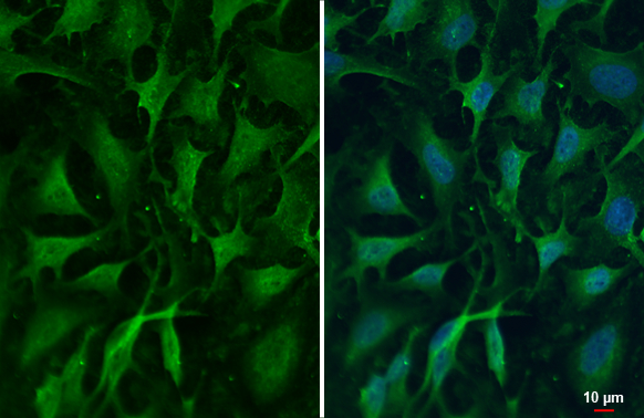
ZC3H12A antibody detects ZC3H12A protein at cytoplasm by immunofluorescent analysis. Sample: HeLa cells were fixed in 4% paraformaldehyde at RT for 15 min. Green: ZC3H12A stained by ZC3H12A antibody (GTX110807) diluted at 1:500.
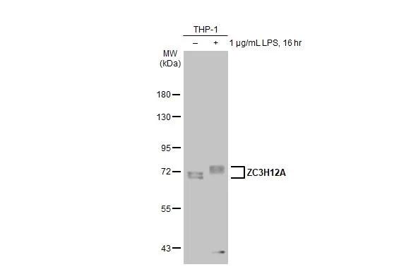
![Untreated (–) and treated (+) THP-1 whole cell extracts (30 μg) were separated by 7.5% SDS-PAGE, and the membrane was blotted with ZC3H12A antibody [N3C3] (GTX110807) diluted at 1:1000. The HRP-conjugated anti-rabbit IgG antibody (GTX213110-01) was used to detect the primary antibody.](https://www.genetex.com/upload/website/prouct_img/normal/GTX110807/GTX110807_44692_20220610_WB_treatment_LPS_24062501_442.webp "Untreated (–) and treated (+) THP-1 whole cell extracts (30 μg) were separated by 7.5% SDS-PAGE, and the membrane was blotted with ZC3H12A antibody [N3C3] (GTX110807) diluted at 1:1000. The HRP-conjugated anti-rabbit IgG antibody (GTX213110-01) was used to detect the primary antibody.")
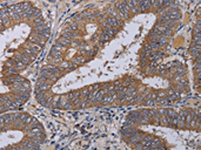
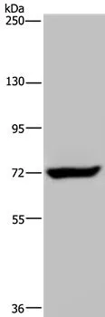
![Various tissue extracts (30 μg) were separated by 7.5% SDS-PAGE, and the membrane was blotted with ZC3H12A antibody [N3C3] (GTX110807) diluted at 1:1000. The HRP-conjugated anti-rabbit IgG antibody (GTX213110-01) was used to detect the primary antibody, and the signal was developed with Trident ECL plus-Enhanced.](https://www.genetex.com/upload/website/prouct_img/normal/GTX110807/GTX110807_44692_20220729_WB_M_tissue_24062501_167.webp "Various tissue extracts (30 μg) were separated by 7.5% SDS-PAGE, and the membrane was blotted with ZC3H12A antibody [N3C3] (GTX110807) diluted at 1:1000. The HRP-conjugated anti-rabbit IgG antibody (GTX213110-01) was used to detect the primary antibody, and the signal was developed with Trident ECL plus-Enhanced.")
ZC3H12A antibody detects ZC3H12A protein at cytoplasm by immunofluorescent analysis. Sample: HeLa cells were fixed in 4% paraformaldehyde at RT for 15 min. Green: ZC3H12A stained by ZC3H12A antibody (GTX110807) diluted at 1:500.
ZC3H12A antibody [N3C3]
GTX110807
ApplicationsFlow Cytometry, ImmunoFluorescence, ImmunoPrecipitation, Western Blot, ImmunoCytoChemistry, ImmunoHistoChemistry
Product group Antibodies
ReactivityHuman, Mouse
TargetZC3H12A
Overview
- SupplierGeneTex
- Product NameZC3H12A antibody [N3C3]
- Delivery Days Customer9
- Application Supplier NoteWB: 1:500-1:3000. ICC/IF: 1:100-1:1000. *Optimal dilutions/concentrations should be determined by the researcher.Not tested in other applications.
- ApplicationsFlow Cytometry, ImmunoFluorescence, ImmunoPrecipitation, Western Blot, ImmunoCytoChemistry, ImmunoHistoChemistry
- CertificationResearch Use Only
- ClonalityPolyclonal
- Concentration0.5 mg/ml
- ConjugateUnconjugated
- Gene ID80149
- Target nameZC3H12A
- Target descriptionzinc finger CCCH-type containing 12A
- Target synonymsMCPIP, MCPIP-1, MCPIP1, Reg1, dJ423B22.1, endoribonuclease ZC3H12A, MCP-1 treatment-induced protein, MCP-induced protein 1, bifunctional endoribonuclease and deubiquitinase ZC3H12A, monocyte chemotactic protein-induced protein 1, regnase-1, ribonuclease ZC3H12A, zinc finger CCCH domain-containing protein 12A
- HostRabbit
- IsotypeIgG
- Protein IDQ5D1E8
- Protein NameEndoribonuclease ZC3H12A
- Scientific DescriptionZC3H12A is an MCP1 (CCL2; MIM 158105)-induced protein that acts as a transcriptional activator and causes cell death of cardiomyocytes, possibly via induction of genes associated with apoptosis.[supplied by OMIM]
- ReactivityHuman, Mouse
- Storage Instruction-20°C or -80°C,2°C to 8°C
- UNSPSC41116161
Datasheet
Related products

Product group Antibodies
Anti-ZC3H12A AntibodyA36858
ApplicationsWestern Blot, ImmunoHistoChemistry
ReactivityHuman
- SizePrice

Product group Antibodies
Anti-MCPIP1/ZC3H12A Antibody Picoband(r)A01688-2-CARRIER-FREE
ApplicationsFlow Cytometry, ImmunoFluorescence, Western Blot, ELISA, ImmunoCytoChemistry, ImmunoHistoChemistry
ReactivityHuman, Mouse, Rat
TargetZC3H12A
- SizePrice
Product group Antibodies
MCPIP1 Polyclonal Antibodybs-5792R
ApplicationsFlow Cytometry, ImmunoFluorescence, Western Blot, ELISA, ImmunoCytoChemistry, ImmunoHistoChemistry, ImmunoHistoChemistry Frozen, ImmunoHistoChemistry Paraffin
ReactivityBovine, Canine, Chicken, Equine, Human, Mouse, Rabbit, Rat
TargetZC3H12A
- SizePrice
Product group Antibodies
Zc3H12A Polyclonal AntibodyCAC10457
ApplicationsELISA, ImmunoHistoChemistry
TargetZC3H12A
- SizePrice
Product group Antibodies
ZC3H12A AntibodyCSB-PA037047
ApplicationsWestern Blot, ELISA, ImmunoHistoChemistry
ReactivityHuman
TargetZC3H12A
- SizePrice
Product group Antibodies
ZC3H12A antibodyGTX02811
ApplicationsWestern Blot, ImmunoHistoChemistry, ImmunoHistoChemistry Paraffin
ReactivityHuman
TargetZC3H12A
- SizePrice
Product group Antibodies
ZC3H12A antibodyGTX110808
ApplicationsWestern Blot
ReactivityHuman
TargetZC3H12A
- SizePrice
![Non-transfected (–) and transfected (+) 293T whole cell extracts (30 μg) were separated by 7.5% SDS-PAGE, and the membrane was blotted with ZC3H12A antibody [HL1442] (GTX636914) diluted at 1:1000. The HRP-conjugated anti-rabbit IgG antibody (GTX213110-01) was used to detect the primary antibody, and the signal was developed with Trident ECL plus-Enhanced.](https://www.genetex.com/upload/website/prouct_img/normal/GTX636914/GTX636914_44697_20220819_WB_shRNA_watermark_22082402_798.webp)
Product group Antibodies
ZC3H12A antibody [HL1442]GTX636914
ApplicationsWestern Blot
ReactivityFeline, Human
TargetZC3H12A
- SizePrice

Product group Antibodies
Anti-ZC3H12A AntibodyHPA032052
ApplicationsImmunoHistoChemistry
ReactivityHuman
TargetZC3H12A
- SizePrice