

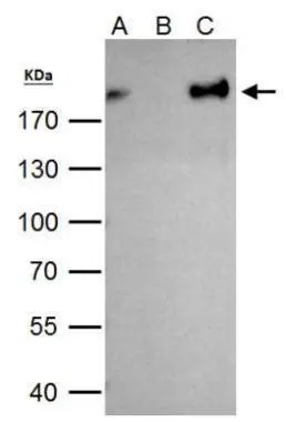
ZEB1 antibody immunoprecipitates ZEB1 protein in IP experiments. IP Sample: 293T whole cell lysate/extract A. 40 μg 293T whole cell lysate/extract B. Control with 2 μg of preimmune rabbit IgG C. Immunoprecipitation of ZEB1 protein by 2 μg of ZEB1 antibody (GTX105278) 7.5% SDS-PAGE The immunoprecipitated ZEB1 protein was detected by ZEB1 antibody (GTX105278) diluted at 1:1000. EasyBlot anti-rabbit IgG (GTX221666-01) was used as a secondary reagent.

![ZEB1 antibody [N2C1], Internal detects ZEB1 protein at nucleus by immunofluorescent analysis. Sample: HeLa cells were fixed in 4% paraformaldehyde at RT for 15 min. Green: ZEB1 protein stained by ZEB1 antibody [N2C1], Internal (GTX105278) diluted at 1:500. Red: Phalloidin, a cytoskeleton marker, diluted at 1:100. Scale bar = 10 μm.](https://www.genetex.com/upload/website/prouct_img/normal/GTX105278/GTX105278_42732_20170315_IFA_w_23060120_193.webp "ZEB1 antibody [N2C1], Internal detects ZEB1 protein at nucleus by immunofluorescent analysis. Sample: HeLa cells were fixed in 4% paraformaldehyde at RT for 15 min. Green: ZEB1 protein stained by ZEB1 antibody [N2C1], Internal (GTX105278) diluted at 1:500. Red: Phalloidin, a cytoskeleton marker, diluted at 1:100. Scale bar = 10 μm.")
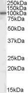
![Various whole cell extracts (30 μg) were separated by 5% SDS-PAGE, and the membranes were blotted with ZEB1 antibody [N2C1], Internal (GTX105278) diluted at 1:1000 and competitor's antibody diluted at 1:1000. The HRP-conjugated anti-rabbit IgG antibody (GTX213110-01) was used to detect the primary antibody. *The competitor is not affiliated with GeneTex and does not endorse this product.](https://www.genetex.com/upload/website/prouct_img/normal/GTX105278/GTX105278_42690_20200403_WB_competitor_watermark_w_23060120_328.webp "Various whole cell extracts (30 μg) were separated by 5% SDS-PAGE, and the membranes were blotted with ZEB1 antibody [N2C1], Internal (GTX105278) diluted at 1:1000 and competitor's antibody diluted at 1:1000. The HRP-conjugated anti-rabbit IgG antibody (GTX213110-01) was used to detect the primary antibody. *The competitor is not affiliated with GeneTex and does not endorse this product.")
![Non-transfected (–) and transfected (+) HeLa whole cell extracts (30 μg) were separated by 5% SDS-PAGE, and the membrane was blotted with ZEB1 antibody [N2C1], Internal (GTX105278) diluted at 1:500. The HRP-conjugated anti-rabbit IgG antibody (GTX213110-01) was used to detect the primary antibody, and the signal was developed with Trident ECL plus-Enhanced.](https://www.genetex.com/upload/website/prouct_img/normal/GTX105278/GTX105278_42690_20180810_WB_shRNA_watermark_w_23060120_479.webp "Non-transfected (–) and transfected (+) HeLa whole cell extracts (30 μg) were separated by 5% SDS-PAGE, and the membrane was blotted with ZEB1 antibody [N2C1], Internal (GTX105278) diluted at 1:500. The HRP-conjugated anti-rabbit IgG antibody (GTX213110-01) was used to detect the primary antibody, and the signal was developed with Trident ECL plus-Enhanced.")
 were separated by 5% SDS-PAGE, and the membrane was blotted with ZEB1 antibody (GTX105278) diluted at 1:1000. The HRP-conjugated anti-rabbit IgG antibody (GTX213110-01) was used to detect the primary antibody.")
.")
![Various whole cell extracts (30 μg) were separated by 5% SDS-PAGE, and the membrane was blotted with ZEB1 antibody [N2C1], Internal (GTX105278) diluted at 1:1000. The HRP-conjugated anti-rabbit IgG antibody (GTX213110-01) was used to detect the primary antibody. Corresponding RNA expression data for the same cell lines are based on Human Protein Atlas program.](https://www.genetex.com/upload/website/prouct_img/normal/GTX105278/GTX105278_44972_20230317_WB_TPM_watermark_24062501_365.webp "Various whole cell extracts (30 μg) were separated by 5% SDS-PAGE, and the membrane was blotted with ZEB1 antibody [N2C1], Internal (GTX105278) diluted at 1:1000. The HRP-conjugated anti-rabbit IgG antibody (GTX213110-01) was used to detect the primary antibody. Corresponding RNA expression data for the same cell lines are based on Human Protein Atlas program.")
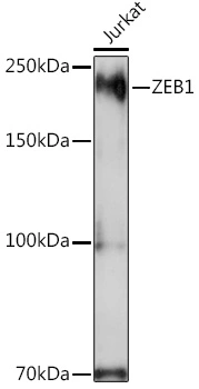
![Various whole cell extracts (30 μg) were separated by 5% SDS-PAGE, and the membrane was blotted with ZEB1 antibody [N2C1], Internal (GTX105278) diluted at 1:1000. The HRP-conjugated anti-rabbit IgG antibody (GTX213110-01) was used to detect the primary antibody.](https://www.genetex.com/upload/website/prouct_img/normal/GTX105278/GTX105278_44972_20230303_WB_M_R_24062501_183.webp "Various whole cell extracts (30 μg) were separated by 5% SDS-PAGE, and the membrane was blotted with ZEB1 antibody [N2C1], Internal (GTX105278) diluted at 1:1000. The HRP-conjugated anti-rabbit IgG antibody (GTX213110-01) was used to detect the primary antibody.")
ZEB1 antibody immunoprecipitates ZEB1 protein in IP experiments. IP Sample: 293T whole cell lysate/extract A. 40 μg 293T whole cell lysate/extract B. Control with 2 μg of preimmune rabbit IgG C. Immunoprecipitation of ZEB1 protein by 2 μg of ZEB1 antibody (GTX105278) 7.5% SDS-PAGE The immunoprecipitated ZEB1 protein was detected by ZEB1 antibody (GTX105278) diluted at 1:1000. EasyBlot anti-rabbit IgG (GTX221666-01) was used as a secondary reagent.
ZEB1 antibody [N2C1], Internal
GTX105278
ApplicationsImmunoFluorescence, ImmunoPrecipitation, Western Blot, ChIP Chromatin ImmunoPrecipitation, ImmunoCytoChemistry
Product group Antibodies
ReactivityHuman, Mouse, Rat
TargetZEB1
Overview
- SupplierGeneTex
- Product NameZEB1 antibody [N2C1], Internal
- Delivery Days Customer9
- Application Supplier NoteWB: 1:500-1:3000. ICC/IF: 1:100-1:1000. IP: 1:100-1:500. *Optimal dilutions/concentrations should be determined by the researcher.Not tested in other applications.
- ApplicationsImmunoFluorescence, ImmunoPrecipitation, Western Blot, ChIP Chromatin ImmunoPrecipitation, ImmunoCytoChemistry
- CertificationResearch Use Only
- ClonalityPolyclonal
- Concentration0.13 mg/ml
- ConjugateUnconjugated
- Gene ID6935
- Target nameZEB1
- Target descriptionzinc finger E-box binding homeobox 1
- Target synonymsAREB6, BZP, DELTAEF1, FECD6, NIL2A, PPCD3, TCF8, ZFHEP, ZFHX1A, zinc finger E-box-binding homeobox 1, delta-crystallin enhancer binding factor 1, negative regulator of IL2, posterior polymorphous corneal dystrophy 3, transcription factor 8 (represses interleukin 2 expression), zinc finger homeodomain enhancer-binding protein
- HostRabbit
- IsotypeIgG
- Protein IDP37275
- Protein NameZinc finger E-box-binding homeobox 1
- Scientific DescriptionZEB1 encodes a zinc finger transcription factor that represses T-lymphocyte-specific IL2 gene (MIM 147680) expression by binding to a negative regulatory domain 100 nucleotides 5-prime of the IL2 transcription start site (Williams et al., 1991 [PubMed 1840704]).[supplied by OMIM]
- ReactivityHuman, Mouse, Rat
- Storage Instruction-20°C or -80°C,2°C to 8°C
- UNSPSC12352203
References
- Hung TH, Huang Y, Yeh CT, et al. High expression of embryonic stem cell marker SSEA3 confers poor prognosis and promotes epithelial mesenchymal transition in hepatocellular carcinoma. Biomed J. 2024,47(2):100612. doi: 10.1016/j.bj.2023.100612Read this paper
- Hsu PL, Chien CW, Tang YA, et al. Targeting BRD3 eradicates nuclear TYRO3-induced colorectal cancer metastasis. Sci Adv. 2023,9(15):eade3422. doi: 10.1126/sciadv.ade3422Read this paper
- Chiou WC, Huang GJ, Chang TY, et al. Ovatodiolide inhibits SARS-CoV-2 replication and ameliorates pulmonary fibrosis through suppression of the TGF-β/TβRs signaling pathway. Biomed Pharmacother. 2023,161:114481. doi: 10.1016/j.biopha.2023.114481Read this paper
- Wuputra K, Hsiao PJ, Chang WT, et al. FOXM1-CD44 Signaling Is Critical for the Acquisition of Regorafenib Resistance in Human Liver Cancer Cells. Int J Mol Sci. 2022,23(14). doi: 10.3390/ijms23147782Read this paper
- Plaschka M, Benboubker V, Grimont M, et al. ZEB1 transcription factor promotes immune escape in melanoma. J Immunother Cancer. 2022,10(3). doi: 10.1136/jitc-2021-003484Read this paper
- Tsai SC, Wu WC, Yang JS. Tetrandrine Inhibits Epithelial-Mesenchymal Transition in IL-6-Induced HCT116 Human Colorectal Cancer Cells. Onco Targets Ther. 2021,14:4523-4536. doi: 10.2147/OTT.S324552Read this paper
- Li JN, Sun HL, Wang MY, et al. E-cadherin Interacts With Posttranslationally-Modified AGO2 to Enhance miRISC Activity. Front Cell Dev Biol. 2021,9:671244. doi: 10.3389/fcell.2021.671244Read this paper
- Wang YY, Chen HD, Lo S, et al. Visfatin Enhances Breast Cancer Progression through CXCL1 Induction in Tumor-Associated Macrophages. Cancers (Basel). 2020,12(12). doi: 10.3390/cancers12123526Read this paper
- Li CL, Huang CW, Ko CJ, et al. Curcumol Suppresses Triple-negative Breast Cancer Metastasis by Attenuating Anoikis Resistance via Inhibition of Skp2-mediated Transcriptional Addiction. Anticancer Res. 2020,40(10):5529-5538. doi: 10.21873/anticanres.14565Read this paper
- Lee JY, Fan CC, Chou NL, et al. PHRF1 promotes migration and invasion by modulating ZEB1 expression. PLoS One. 2020,15(7):e0236876. doi: 10.1371/journal.pone.0236876Read this paper
Datasheet
Related products
Product group Antibodies

Anti-ZEB1 [RAB-T19]Ab01935-1.1
ApplicationsFlow Cytometry, ImmunoFluorescence
ReactivityHuman
TargetZEB1
- SizePrice
Product group Antibodies
Anti-ZEB1 Antibody144-63542
ApplicationsImmunoFluorescence, Western Blot, ImmunoHistoChemistry
ReactivityHuman, Mouse, Rat
TargetZEB1
- SizePrice

Product group Antibodies
Anti-AREB6/ZEB1 Antibody Picoband(r)A00548-2-CARRIER-FREE
ApplicationsFlow Cytometry, ImmunoFluorescence, Western Blot, ImmunoCytoChemistry, ImmunoHistoChemistry
ReactivityHuman
TargetZEB1
- SizePrice
![Whole cell extract (30 μg) was separated by 5% SDS-PAGE, and the membrane was blotted with ZEB1 antibody [HL2245] (GTX638294) diluted at 1:1000. The HRP-conjugated anti-rabbit IgG antibody (GTX213110-01) was used to detect the primary antibody.](https://www.genetex.com/upload/website/prouct_img/normal/GTX638294/GTX638294_T-44960_20230224_WB_M_23030219_377.webp)
Product group Antibodies
ZEB1 antibody [HL2245]GTX638294
ApplicationsImmunoFluorescence, Western Blot, ImmunoCytoChemistry, ImmunoHistoChemistry, ImmunoHistoChemistry Paraffin
ReactivityHuman, Mouse, Rat
TargetZEB1
- SizePrice
Product group Antibodies
ApplicationsImmunoPrecipitation, Western Blot, ImmunoCytoChemistry, ImmunoHistoChemistry
TargetZEB1
- SizePrice

Product group Antibodies
Anti-ZEB1 AntibodyA13473
ApplicationsImmunoFluorescence, Western Blot, ImmunoCytoChemistry, ImmunoHistoChemistry
ReactivityHuman, Mouse, Rat
- SizePrice
Product group Antibodies
ZEB1 antibody, InternalGTX88467
ApplicationsWestern Blot
ReactivityHuman, Mouse
TargetZEB1
- SizePrice
Product group Antibodies
References
ZEB1 antibodyGTX33589
ApplicationsImmunoFluorescence, Western Blot, ImmunoCytoChemistry, ImmunoHistoChemistry, ImmunoHistoChemistry Paraffin
ReactivityHuman, Mouse, Rat
TargetZEB1
- SizePrice
Product group Antibodies
References
ZEB1 antibodyGTX55847
ApplicationsWestern Blot, ImmunoHistoChemistry, ImmunoHistoChemistry Paraffin
ReactivityHuman
TargetZEB1
- SizePrice