

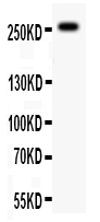
WB analysis of 50ug mouse brain tissue lysate using GTX12302 ABCA4 antibody. Dilution : 0.5 μg/mL
WB analysis of 50ug mouse brain tissue lysate using GTX12302 ABCA4 antibody. Dilution : 0.5 μg/mL
ABCA4 antibody
GTX12302
ApplicationsWestern Blot
Product group Antibodies
ReactivityMouse, Rat
TargetAbca4
Overview
- SupplierGeneTex
- Product NameABCA4 antibody
- Delivery Days Customer9
- Application Supplier NoteWB: 0.1-0.5microg/ml. *Optimal dilutions/concentrations should be determined by the researcher.Not tested in other applications.
- ApplicationsWestern Blot
- CertificationResearch Use Only
- ClonalityPolyclonal
- Concentration500 ug/ml
- ConjugateUnconjugated
- Gene ID11304
- Target nameAbca4
- Target descriptionATP-binding cassette, sub-family A member 4
- Target synonymsAbc10, Abcr, D430003I15Rik, RmP, retinal-specific phospholipid-transporting ATPase ABCA4, ATP-binding cassette 10, ATP-binding cassette, sub-family A (ABC1), member 4, RIM ABC transporter, Rim protein, retinal-specific ATP-binding cassette transporter
- HostRabbit
- IsotypeIgG
- Protein IDO35600
- Protein NameRetinal-specific phospholipid-transporting ATPase ABCA4
- Scientific DescriptionThe membrane-associated protein encoded by this gene is a member of the superfamily of ATP-binding cassette (ABC) transporters. ABC proteins transport various molecules across extra- and intracellular membranes. ABC genes are divided into seven distinct subfamilies (ABC1, MDR/TAP, MRP, ALD, OABP, GCN20, White). This protein is a member of the ABC1 subfamily. Members of the ABC1 subfamily comprise the only major ABC subfamily found exclusively in multicellular eukaryotes. This protein was the first of the ABC transporters to be observed in photoreceptors and may play a role in the photoresponse. Mutations in the human gene are found in patients diagnosed with Stargardt disease and are associated with retinitis pigmentosa-19 and macular degeneration age-related 2. [provided by RefSeq, Jul 2008]
- ReactivityMouse, Rat
- Storage Instruction-20°C or -80°C,2°C to 8°C
- UNSPSC12352203
Datasheet
Related products
Product group Antibodies
ABCA4 (Rim Protein) AntibodyBSM-70010M
ApplicationsWestern Blot, ImmunoHistoChemistry, ImmunoHistoChemistry Paraffin
ReactivityBovine, Human, Mouse, Xenopus
TargetAbca4
- SizePrice

Product group Antibodies
Anti-ABCA4 Antibody Picoband(r)PA2066-CARRIER-FREE
ApplicationsWestern Blot
ReactivityMouse
TargetAbca4
- SizePrice
![Immunohistochemical staining of adult mouse retina showing specific immunolabeling of the ABCA4 protein using ABCA4 (Rim Protein) antibody [3F4] (GTX82741).](https://www.genetex.com/upload/website/prouct_img/normal/GTX82741/ABCA4-Rim-Protein-antibody-3F4_Immunohistochemistry_GTX82741-1_w_23061322_724.webp)
Product group Antibodies
References
ApplicationsWestern Blot, ImmunoHistoChemistry
ReactivityBovine, Human, Mouse, Xenopus
TargetAbca4
- SizePrice