

Immunohistochemistry of paraffin-embedded Human brain tissue using ABL1 Polyclonal Antibody at dilution 1:40
Immunohistochemistry of paraffin-embedded Human brain tissue using ABL1 Polyclonal Antibody at dilution 1:40
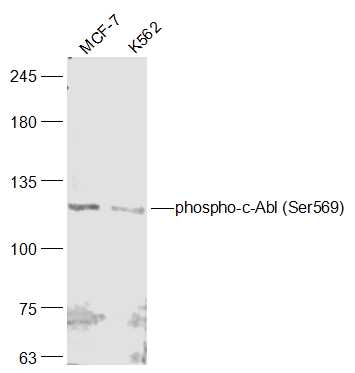
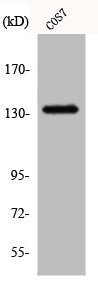
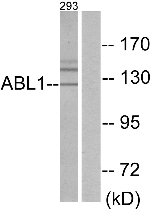
ABL1 Polyclonal Antibody
E-AB-10119
Product group Antibodies
Overview
- SupplierElabscience
- Product NameABL1 Polyclonal Antibody
- Delivery Days Customer12
- Applications SupplierELISA IHC
- CertificationResearch Use Only
- Concentration0.5mg/ml
- Scientific DescriptionThe ABL1 protooncogene encodes a cytoplasmic and nuclear protein tyrosine kinase that has been implicated in processes of cell differentiation, cell division, cell adhesion, and stress response. Activity of c-Abl protein is negatively regulated by its SH3 domain, and deletion of the SH3 domain turns ABL1 into an oncogene. The t(9;22) translocation results in the head-to-tail fusion of the BCR (MIM:151410) and ABL1 genes present in many cases of chronic myelogeneous leukemia. The DNA-binding activity of the ubiquitously expressed ABL1 tyrosine kinase is regulated by CDC2-mediated phosphorylation, suggesting a cell cycle function for ABL1.
- UNSPSC12352203
Related products
Product group Antibodies
ABL1 Polyclonal AntibodyCAC14060
ApplicationsImmunoPrecipitation, ELISA, ImmunoHistoChemistry
TargetABL1
- SizePrice
Product group Antibodies
ApplicationsImmunoFluorescence, Western Blot, ELISA, ImmunoCytoChemistry, ImmunoHistoChemistry, ImmunoHistoChemistry Frozen, ImmunoHistoChemistry Paraffin
TargetABL1
- SizePrice
Product group Antibodies
References
c-Abl antibodyGTX111317
ApplicationsImmunoFluorescence, Western Blot, ImmunoCytoChemistry, ImmunoHistoChemistry, ImmunoHistoChemistry Paraffin
TargetABL1
- SizePrice
Product group Antibodies
ABL1 AntibodyCSB-PA001140
ApplicationsWestern Blot, ELISA, ImmunoHistoChemistry
TargetABL1
- SizePrice
Product group Antibodies
ApplicationsImmunoFluorescence, ImmunoPrecipitation, Western Blot, ImmunoCytoChemistry, ImmunoHistoChemistry
TargetABL1
- SizePrice
Product group Antibodies
Anti-ABL1 AntibodyA96250
ApplicationsWestern Blot, ELISA, ImmunoHistoChemistry
- SizePrice
Product group Antibodies
Anti-ABL1 Antibody144-00282
ApplicationsImmunoFluorescence, Western Blot, ImmunoHistoChemistry
TargetABL1
- SizePrice

Product group Antibodies
Anti-ABL1 Antibody Picoband(r)A00133-1-CARRIER-FREE
ApplicationsFlow Cytometry, ImmunoFluorescence, Western Blot, ELISA, ImmunoCytoChemistry
TargetABL1
- SizePrice