

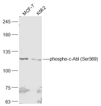
Lane 1: MCF-7 lysates; Lane 2: K562 lysates probed with c-Abl (Ser569) Polyclonal Antibody, Unconjugated (bs-12904R) at 1:300 dilution and 4˚C overnight incubation. Followed by conjugated secondary antibody incubation at 1:10000 for 60 min at 37˚C.
Lane 1: MCF-7 lysates; Lane 2: K562 lysates probed with c-Abl (Ser569) Polyclonal Antibody, Unconjugated (bs-12904R) at 1:300 dilution and 4˚C overnight incubation. Followed by conjugated secondary antibody incubation at 1:10000 for 60 min at 37˚C.
phospho-c-Abl (Ser569) Polyclonal Antibody
BS-12904R
ApplicationsImmunoFluorescence, Western Blot, ELISA, ImmunoCytoChemistry, ImmunoHistoChemistry, ImmunoHistoChemistry Frozen, ImmunoHistoChemistry Paraffin
Product group Antibodies
ReactivityBovine, Equine, Human, Porcine, Sheep
TargetABL1
Overview
- SupplierBioss
- Product Namephospho-c-Abl (Ser569) Polyclonal Antibody
- Delivery Days Customer16
- ApplicationsImmunoFluorescence, Western Blot, ELISA, ImmunoCytoChemistry, ImmunoHistoChemistry, ImmunoHistoChemistry Frozen, ImmunoHistoChemistry Paraffin
- Applications SupplierWB(1:300-5000), ELISA(1:500-1000), IHC-P(1:200-400), IHC-F(1:100-500), IF(IHC-P)(1:50-200), IF(IHC-F)(1:50-200), IF(ICC)(1:50-200)
- CertificationResearch Use Only
- ClonalityPolyclonal
- Concentration1 ug/ul
- ConjugateUnconjugated
- Gene ID25
- Target nameABL1
- Target descriptionABL proto-oncogene 1, non-receptor tyrosine kinase
- Target synonymsABL, BCR-ABL, CHDSKM, JTK7, bcr/abl, c-ABL, c-ABL1, p150, v-abl, tyrosine-protein kinase ABL1, ABL protooncogene 1 nonreceptor tyrosine kinase, Abelson tyrosine-protein kinase 1, BCR-ABL1 p190, BCR/ABL e8a2 fusion, BCR/ABL1 e1a2 fusion protein, BCR/ABL1 fusion, BCR/ABL1 fusion protein e3a1, BCR::ABL1 fusion protein, bcr/c-abl oncogene protein, c-abl oncogene 1, receptor tyrosine kinase, chimeric BCR::ABL1 protein, proto-oncogene c-Abl, proto-oncogene tyrosine-protein kinase ABL1, v-abl Abelson murine leukemia viral oncogene homolog 1
- HostRabbit
- IsotypeIgG
- ReactivityBovine, Equine, Human, Porcine, Sheep
- Storage Instruction-20°C
- UNSPSC41116161
Datasheet
Related products
Product group Antibodies
Anti-ABL1 AntibodyA96250
ApplicationsWestern Blot, ELISA, ImmunoHistoChemistry
ReactivityHuman, Mouse, Rat
- SizePrice
Product group Antibodies
Anti-ABL1 Antibody144-00282
ApplicationsImmunoFluorescence, Western Blot, ImmunoHistoChemistry
ReactivityHuman, Mouse, Rat
TargetABL1
- SizePrice

Product group Antibodies
Anti-ABL1 Antibody Picoband(r)A00133-1-CARRIER-FREE
ApplicationsFlow Cytometry, ImmunoFluorescence, Western Blot, ELISA, ImmunoCytoChemistry
ReactivityHuman, Mouse, Rat
TargetABL1
- SizePrice
Product group Antibodies
ABL1 AntibodyCSB-PA001140
ApplicationsWestern Blot, ELISA, ImmunoHistoChemistry
ReactivityHuman, Monkey, Mouse, Rat
TargetABL1
- SizePrice
Product group Antibodies
ABL1 Polyclonal AntibodyCAC14060
ApplicationsImmunoPrecipitation, ELISA, ImmunoHistoChemistry
TargetABL1
- SizePrice

Product group Antibodies
Anti-ABL1 AntibodyHPA027251
ApplicationsImmunoHistoChemistry
ReactivityHuman
TargetABL1
- SizePrice
Product group Antibodies
c-Abl antibodyGTX111317
ApplicationsImmunoFluorescence, Western Blot, ImmunoCytoChemistry, ImmunoHistoChemistry, ImmunoHistoChemistry Paraffin
ReactivityHuman
TargetABL1
- SizePrice
Product group Antibodies

ABL1 / c-ABL AntibodyLS-C400536
ApplicationsELISA, ImmunoHistoChemistry
ReactivityHuman, Mouse
TargetABL1
- SizePrice
Product group Antibodies
Anti-ABL1 AntibodyCAB0282
ApplicationsImmunoFluorescence, Western Blot, ELISA, ImmunoCytoChemistry, ImmunoHistoChemistry, ImmunoHistoChemistry Paraffin
ReactivityHuman
TargetABL1
- SizePrice