


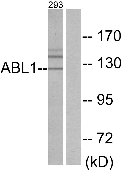
Figure 1. Western blot analysis of ABL1 using anti-ABL1 antibody (A00133-1). Electrophoresis was performed on a 5-20% SDS-PAGE gel at 70V (Stacking gel) / 90V (Resolving gel) for 2-3 hours. The sample well of each lane was loaded with 30 ug of sample under reducing conditions. Lane 1: human Jurkat whole cell lysates, Lane 2: human MCF-7 whole cell lysates, Lane 3: human MDA-MB-453 whole cell lysates, Lane 4: human Raji whole cell lysates. After electrophoresis, proteins were transferred to a nitrocellulose membrane at 150 mA for 50-90 minutes. Blocked the membrane with 5% non-fat milk/TBS for 1.5 hour at RT. The membrane was incubated with rabbit anti-ABL1 antigen affinity purified polyclonal antibody (Catalog # A00133-1) at 0.5 microg/mL overnight at 4°C, then washed with TBS-0.1%Tween 3 times with 5 minutes each and probed with a goat anti-rabbit IgG-HRP secondary antibody at a dilution of 1:5000 for 1.5 hour at RT. The signal is developed using an Enhanced Chemiluminescent detection (ECL) kit (Catalog # EK1002) with Tanon 5200 system. A specific band was detected for ABL1 at approximately 143 kDa. The expected band size for ABL1 is at 133 kDa.
 and anti-Beta Tubulin antibody (M01857-3). ABL1 was detected in immunocytochemical section of U2OS cell. Enzyme antigen retrieval was performed using IHC enzyme antigen retrieval reagent (AR0022) for 15 mins. The cells were blocked with 10% goat serum. And then incubated with 5 microg/mL rabbit anti-ABL1 Antibody (A00133-1) and mouse anti-Beta Tubulin antibody (M01857-3) overnight at 4°C. DyLight®488 Conjugated Goat Anti-Rabbit IgG (BA1127) and DyLight®550 Conjugated Goat Anti-Mouse IgG (BA1133) were used as secondary antibody at 1:500 dilution and incubated for 30 minutes at 37°C. Visualize using a fluorescence microscope and filter sets appropriate for the label used.")
. Overlay histogram showing PC-3 cells stained with A00133-1 (Blue line). To facilitate intracellular staining, cells were fixed with 4% paraformaldehyde and permeabilized with permeabilization buffer. The cells were blocked with 10% normal goat serum. And then incubated with rabbit anti-ABL1 Antibody (A00133-1, 1 microg/1x106 cells) for 30 min at 20°C. DyLight®488 conjugated goat anti-rabbit IgG (BA1127, 5-10 microg/1x106 cells) was used as secondary antibody for 30 minutes at 20°C. Isotype control antibody (Green line) was rabbit IgG (1 microg/1x106) used under the same conditions. Unlabelled sample (Red line) was also used as a control.")
. Overlay histogram showing PC-12 cells stained with A00133-1 (Blue line). To facilitate intracellular staining, cells were fixed with 4% paraformaldehyde and permeabilized with permeabilization buffer. The cells were blocked with 10% normal goat serum. And then incubated with rabbit anti-ABL1 Antibody (A00133-1, 1 microg/1x106 cells) for 30 min at 20°C. DyLight®488 conjugated goat anti-rabbit IgG (BA1127, 5-10 microg/1x106 cells) was used as secondary antibody for 30 minutes at 20°C. Isotype control antibody (Green line) was rabbit IgG (1 microg/1x106) used under the same conditions. Unlabelled sample (Red line) was also used as a control.")
. Overlay histogram showing RAW264.7 cells stained with A00133-1 (Blue line). To facilitate intracellular staining, cells were fixed with 4% paraformaldehyde and permeabilized with permeabilization buffer. The cells were blocked with 10% normal goat serum. And then incubated with rabbit anti-ABL1 Antibody (A00133-1, 1 microg/1x106 cells) for 30 min at 20°C. DyLight®488 conjugated goat anti-rabbit IgG (BA1127, 5-10 microg/1x106 cells) was used as secondary antibody for 30 minutes at 20°C. Isotype control antibody (Green line) was rabbit IgG (1 microg/1x106) used under the same conditions. Unlabelled sample (Red line) was also used as a control.")
Figure 1. Western blot analysis of ABL1 using anti-ABL1 antibody (A00133-1). Electrophoresis was performed on a 5-20% SDS-PAGE gel at 70V (Stacking gel) / 90V (Resolving gel) for 2-3 hours. The sample well of each lane was loaded with 30 ug of sample under reducing conditions. Lane 1: human Jurkat whole cell lysates, Lane 2: human MCF-7 whole cell lysates, Lane 3: human MDA-MB-453 whole cell lysates, Lane 4: human Raji whole cell lysates. After electrophoresis, proteins were transferred to a nitrocellulose membrane at 150 mA for 50-90 minutes. Blocked the membrane with 5% non-fat milk/TBS for 1.5 hour at RT. The membrane was incubated with rabbit anti-ABL1 antigen affinity purified polyclonal antibody (Catalog # A00133-1) at 0.5 microg/mL overnight at 4°C, then washed with TBS-0.1%Tween 3 times with 5 minutes each and probed with a goat anti-rabbit IgG-HRP secondary antibody at a dilution of 1:5000 for 1.5 hour at RT. The signal is developed using an Enhanced Chemiluminescent detection (ECL) kit (Catalog # EK1002) with Tanon 5200 system. A specific band was detected for ABL1 at approximately 143 kDa. The expected band size for ABL1 is at 133 kDa.
Anti-ABL1 Antibody Picoband(r)
A00133-1-CARRIER-FREE
ApplicationsFlow Cytometry, ImmunoFluorescence, Western Blot, ELISA, ImmunoCytoChemistry
Product group Antibodies
ReactivityHuman, Mouse, Rat
TargetABL1
Overview
- SupplierBoster Bio
- Product NameAnti-ABL1 Antibody Picoband(r)
- Delivery Days Customer9
- ApplicationsFlow Cytometry, ImmunoFluorescence, Western Blot, ELISA, ImmunoCytoChemistry
- CertificationResearch Use Only
- ClonalityPolyclonal
- Concentration500 ug/ml
- Gene ID25
- Target nameABL1
- Target descriptionABL proto-oncogene 1, non-receptor tyrosine kinase
- Target synonymsABL, BCR-ABL, CHDSKM, JTK7, bcr/abl, c-ABL, c-ABL1, p150, v-abl, tyrosine-protein kinase ABL1, ABL protooncogene 1 nonreceptor tyrosine kinase, Abelson tyrosine-protein kinase 1, BCR-ABL1 p190, BCR/ABL e8a2 fusion, BCR/ABL1 e1a2 fusion protein, BCR/ABL1 fusion, BCR/ABL1 fusion protein e3a1, BCR::ABL1 fusion protein, bcr/c-abl oncogene protein, c-abl oncogene 1, receptor tyrosine kinase, chimeric BCR::ABL1 protein, proto-oncogene c-Abl, proto-oncogene tyrosine-protein kinase ABL1, v-abl Abelson murine leukemia viral oncogene homolog 1
- HostRabbit
- Protein IDP00519
- Protein NameTyrosine-protein kinase ABL1
- Scientific DescriptionBoster Bio Anti-ABL1 Antibody Picoband® catalog # A00133-1. Tested in WB, ICC/IF, Flow Cytometry, ELISA applications. This antibody reacts with Human, Mouse, Rat. The brand Picoband indicates this is a premium antibody that guarantees superior quality, high affinity, and strong signals with minimal background in Western blot applications. Only our best-performing antibodies are designated as Picoband, ensuring unmatched performance.
- ReactivityHuman, Mouse, Rat
- Storage Instruction-20°C,2°C to 8°C
- UNSPSC12352203
Related products
Product group Antibodies
Anti-ABL1 Antibody144-00282
ApplicationsImmunoFluorescence, Western Blot, ImmunoHistoChemistry
ReactivityHuman, Mouse, Rat
TargetABL1
- SizePrice
Product group Antibodies
Anti-ABL1 AntibodyA96250
ApplicationsWestern Blot, ELISA, ImmunoHistoChemistry
ReactivityHuman, Mouse, Rat
- SizePrice
Product group Antibodies
ABL1 Polyclonal AntibodyCAC14060
ApplicationsImmunoPrecipitation, ELISA, ImmunoHistoChemistry
TargetABL1
- SizePrice
Product group Antibodies
ABL1 AntibodyCSB-PA001140
ApplicationsWestern Blot, ELISA, ImmunoHistoChemistry
ReactivityHuman, Monkey, Mouse, Rat
TargetABL1
- SizePrice
Product group Antibodies
c-Abl antibodyGTX111317
ApplicationsImmunoFluorescence, Western Blot, ImmunoCytoChemistry, ImmunoHistoChemistry, ImmunoHistoChemistry Paraffin
ReactivityHuman
TargetABL1
- SizePrice

Product group Antibodies
Anti-ABL1 AntibodyHPA027251
ApplicationsImmunoHistoChemistry
ReactivityHuman
TargetABL1
- SizePrice
Product group Antibodies

ABL1 / c-ABL AntibodyLS-C400536
ApplicationsELISA, ImmunoHistoChemistry
ReactivityHuman, Mouse
TargetABL1
- SizePrice
Product group Antibodies
ApplicationsImmunoFluorescence, Western Blot, ELISA, ImmunoCytoChemistry, ImmunoHistoChemistry, ImmunoHistoChemistry Frozen, ImmunoHistoChemistry Paraffin
ReactivityBovine, Equine, Human, Porcine, Sheep
TargetABL1
- SizePrice
Product group Antibodies
Anti-ABL1 AntibodyCAB0282
ApplicationsImmunoFluorescence, Western Blot, ELISA, ImmunoCytoChemistry, ImmunoHistoChemistry, ImmunoHistoChemistry Paraffin
ReactivityHuman
- SizePrice