

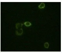
ICC/IF analysis of methanol-fixed HeLa cells using GTX83254 ABL2 antibody [1H1B11]. Green : ABL2
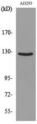
![WB analysis of truncated ABL2 recombinant protein using GTX83254 ABL2 antibody [1H1B11].](https://www.genetex.com/upload/website/prouct_img/normal/GTX83254/GTX83254_20170912_WB_w_23061322_162.webp "WB analysis of truncated ABL2 recombinant protein using GTX83254 ABL2 antibody [1H1B11].")
ICC/IF analysis of methanol-fixed HeLa cells using GTX83254 ABL2 antibody [1H1B11]. Green : ABL2
ABL2 antibody [1H1B11]
GTX83254
ApplicationsImmunoFluorescence, Western Blot, ELISA, ImmunoCytoChemistry
Product group Antibodies
ReactivityHuman
TargetABL2
Overview
- SupplierGeneTex
- Product NameABL2 antibody [1H1B11]
- Delivery Days Customer9
- Application Supplier NoteWB: 1/500 - 1/2000. ICC/IF: 1/200 - 1/1000. ELISA: 1/10000. *Optimal dilutions/concentrations should be determined by the researcher.Not tested in other applications.
- ApplicationsImmunoFluorescence, Western Blot, ELISA, ImmunoCytoChemistry
- CertificationResearch Use Only
- ClonalityMonoclonal
- Clone ID1H1B11
- ConjugateUnconjugated
- Gene ID27
- Target nameABL2
- Target descriptionABL proto-oncogene 2, non-receptor tyrosine kinase
- Target synonymsABLL, ARG, tyrosine-protein kinase ABL2, Abelson tyrosine-protein kinase 2, abelson-related gene protein, c-abl oncogene 2, non-receptor tyrosine kinase, tyrosine-protein kinase ARG, v-abl Abelson murine leukemia viral oncogene homolog 2
- HostMouse
- IsotypeIgG1
- Protein IDP42684
- Protein NameTyrosine-protein kinase ABL2
- Scientific DescriptionThis gene encodes a member of the Abelson family of nonreceptor tyrosine protein kinases. The protein is highly similar to the c-abl oncogene 1 protein, including the tyrosine kinase, SH2 and SH3 domains, and it plays a role in cytoskeletal rearrangements through its C-terminal F-actin- and microtubule-binding sequences. This gene is expressed in both normal and tumor cells, and is involved in translocation with the ets variant 6 gene in leukemia. Multiple alternatively spliced transcript variants encoding different protein isoforms have been found for this gene. [provided by RefSeq, Nov 2009]
- ReactivityHuman
- Storage Instruction-20°C or -80°C,2°C to 8°C
- UNSPSC41116161
Datasheet
Related products
Product group Antibodies
Anti-ABL2 AntibodyA98304
ApplicationsWestern Blot, ELISA
ReactivityHuman, Mouse, Rat
- SizePrice
Product group Antibodies
Anti-ABL2 Antibody102-20129
ApplicationsWestern Blot, ImmunoHistoChemistry, ImmunoHistoChemistry Paraffin
TargetABL2
- SizePrice
Product group Antibodies
ABL2 Recombinant Antibody, Biotin ConjugatedBSM-61381R-BIOTIN
ApplicationsWestern Blot
ReactivityHuman, Mouse, Rat
TargetABL2
- SizePrice
Product group Antibodies
ABL2 AntibodyCSB-PA001106LA01HU
ApplicationsWestern Blot, ELISA
ReactivityHuman
TargetABL2
- SizePrice
Product group Antibodies
ABL2 Polyclonal AntibodyCAC15350
ApplicationsWestern Blot, ELISA
TargetABL2
- SizePrice
Product group Antibodies
ABL2 antibody [N1N3]GTX103584
ApplicationsWestern Blot
ReactivityHuman
TargetABL2
- SizePrice
![WB analysis of HEK293T cells transfected with the pCMV6-ENTRY control (1) and pCMV6-ENTRY ABL2 cDNA (2) using GTX82788 ABL2 antibody [1H1].](https://www.genetex.com/upload/website/prouct_img/normal/GTX82788/GTX82788_20170912_WB_w_23061322_111.webp)
Product group Antibodies
ABL2 antibody [1H1]GTX82788
ApplicationsImmunoFluorescence, Western Blot, ELISA, ImmunoCytoChemistry
ReactivityHuman, Mouse
TargetABL2
- SizePrice

Product group Antibodies
Anti-ABL2 AntibodyHPA072754
ApplicationsImmunoHistoChemistry
ReactivityHuman
TargetABL2
- SizePrice

Product group Antibodies
Anti-ABL2 Antibody Picoband(r)PB9913-CARRIER-FREE
ApplicationsWestern Blot, ImmunoHistoChemistry
ReactivityHuman, Mouse, Rat
TargetABL2
- SizePrice