

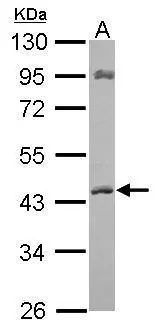
Sample (30 μg of whole cell lysate) A: whole zebrafish 10% SDS PAGE GTX102637 diluted at 1:1000
![ACAT1 antibody [N1N3] detects ACAT1 protein at mitochondria by immunofluorescent analysis. Sample: A431 cells were fixed in 2% paraformaldehyde/culture medium at 37oC for 30 min. Green: ACAT1 protein stained by ACAT1 antibody [N1N3] (GTX102637) diluted at 1:500. Blue: Hoechst 33342 staining.](https://www.genetex.com/upload/website/prouct_img/normal/GTX102637/GTX102637_39764_IFA_w_23060119_560.webp "ACAT1 antibody [N1N3] detects ACAT1 protein at mitochondria by immunofluorescent analysis. Sample: A431 cells were fixed in 2% paraformaldehyde/culture medium at 37oC for 30 min. Green: ACAT1 protein stained by ACAT1 antibody [N1N3] (GTX102637) diluted at 1:500. Blue: Hoechst 33342 staining.")
![ACAT1 antibody [N1N3] detects ACAT1 protein at mitochondria by immunofluorescent analysis. Sample: HeLa cells were fixed in 4% paraformaldehyde at RT for 15 min. Green: ACAT1 protein stained by ACAT1 antibody [N1N3] (GTX102637) diluted at 1:500. Red: alpha Tubulin, a cytoskeleton marker, stained by alpha Tubulin antibody [B-5-1-2] (GTX11304) diluted at 1:10000. Blue: Hoechst 33342 staining.](https://www.genetex.com/upload/website/prouct_img/normal/GTX102637/GTX102637_39764_20150410_IFA_w_23060119_279.webp "ACAT1 antibody [N1N3] detects ACAT1 protein at mitochondria by immunofluorescent analysis. Sample: HeLa cells were fixed in 4% paraformaldehyde at RT for 15 min. Green: ACAT1 protein stained by ACAT1 antibody [N1N3] (GTX102637) diluted at 1:500. Red: alpha Tubulin, a cytoskeleton marker, stained by alpha Tubulin antibody [B-5-1-2] (GTX11304) diluted at 1:10000. Blue: Hoechst 33342 staining.")
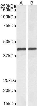
 A: Mouse brain 10% SDS PAGE GTX102637 diluted at 1:1000")
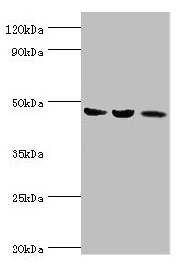
 A:293T B:A431(GTX27909) C:MOLT4(GTX27912) 10% SDS PAGE GTX102637 diluted at 1:500")
![ACAT1 antibody [N1N3] detects ACAT1 protein at cytoplasm on human normal kidney by immunohistochemical analysis. Sample: Paraffin-embedded human normal kidney. ACAT1 antibody [N1N3] (GTX102637) diluted at 1:500.
Antigen Retrieval: Trilogy? (EDTA based, pH 8.0) buffer, 15min](https://www.genetex.com/upload/website/prouct_img/normal/GTX102637/GTX102637_39764_20141128_IHC_w_23060119_573.webp "ACAT1 antibody [N1N3] detects ACAT1 protein at cytoplasm on human normal kidney by immunohistochemical analysis. Sample: Paraffin-embedded human normal kidney. ACAT1 antibody [N1N3] (GTX102637) diluted at 1:500.
Antigen Retrieval: Trilogy? (EDTA based, pH 8.0) buffer, 15min")
Sample (30 μg of whole cell lysate) A: whole zebrafish 10% SDS PAGE GTX102637 diluted at 1:1000
ACAT1 antibody [N1N3]
GTX102637
ApplicationsImmunoFluorescence, Western Blot, ImmunoCytoChemistry, ImmunoHistoChemistry, ImmunoHistoChemistry Paraffin
Product group Antibodies
ReactivityHuman, Mouse, Zebra Fish
TargetACAT1
Overview
- SupplierGeneTex
- Product NameACAT1 antibody [N1N3]
- Delivery Days Customer9
- Application Supplier NoteWB: 1:500-1:3000. ICC/IF: 1:100-1:1000. IHC-P: 1:100-1:1000. *Optimal dilutions/concentrations should be determined by the researcher.Not tested in other applications.
- ApplicationsImmunoFluorescence, Western Blot, ImmunoCytoChemistry, ImmunoHistoChemistry, ImmunoHistoChemistry Paraffin
- CertificationResearch Use Only
- ClonalityPolyclonal
- Concentration1 mg/ml
- ConjugateUnconjugated
- Gene ID38
- Target nameACAT1
- Target descriptionacetyl-CoA acetyltransferase 1
- Target synonymsACAT, MAT, T2, THIL, acetyl-CoA acetyltransferase, mitochondrial, acetoacetyl Coenzyme A thiolase, acetoacetyl-CoA thiolase, acetyl-Coenzyme A acetyltransferase 1, mitochondrial acetoacetyl-CoA thiolase, testicular tissue protein Li 198
- HostRabbit
- IsotypeIgG
- Protein IDP24752
- Protein NameAcetyl-CoA acetyltransferase, mitochondrial
- Scientific DescriptionThis gene encodes a mitochondrially localized enzyme that catalyzes the reversible formation of acetoacetyl-CoA from two molecules of acetyl-CoA. Defects in this gene are associated with 3-ketothiolase deficiency, an inborn error of isoleucine catabolism characterized by urinary excretion of 2-methyl-3-hydroxybutyric acid, 2-methylacetoacetic acid, tiglylglycine, and butanone. [provided by RefSeq]
- ReactivityHuman, Mouse, Zebra Fish
- Storage Instruction-20°C or -80°C,2°C to 8°C
- UNSPSC41116161
Datasheet
Related products
Product group Antibodies
ApplicationsWestern Blot, ELISA
ReactivityHuman, Mouse, Rat
- SizePrice
Product group Antibodies
Anti-ACAT1 Antibody144-60258
ApplicationsWestern Blot, ImmunoHistoChemistry
ReactivityHuman, Mouse
TargetACAT1
- SizePrice
Product group Antibodies

ACAT1 AntibodyLS-C748331
ApplicationsWestern Blot, ImmunoHistoChemistry
ReactivityHuman, Mouse
TargetACAT1
- SizePrice

Product group Antibodies
Anti-ACAT1 Antibody Picoband(r)A02008-1-CARRIER-FREE
ApplicationsFlow Cytometry, ImmunoFluorescence, Western Blot, ELISA, ImmunoCytoChemistry, ImmunoHistoChemistry
ReactivityHuman, Mouse, Rat
TargetACAT1
- SizePrice
Product group Antibodies
ApplicationsFlow Cytometry, Western Blot, ImmunoCytoChemistry
ReactivityHuman, Rat
TargetACAT1
- SizePrice
Product group Antibodies
ACAT1 AntibodyCSB-PA001134ESR1HU
ApplicationsWestern Blot, ELISA, ImmunoHistoChemistry
ReactivityHuman, Mouse, Rat
TargetACAT1
- SizePrice
Product group Antibodies

ApplicationsImmunoFluorescence, Western Blot, ELISA
ReactivityBovine, Human, Mouse, Rat
TargetACAT1
- SizePrice
Product group Antibodies
ACAT1 Polyclonal AntibodyCAC14809
ApplicationsWestern Blot, ELISA, ImmunoHistoChemistry
ReactivityMouse, Rat
TargetACAT1
- SizePrice
Product group Antibodies
ACAT1 antibodyGTX30683
ApplicationsImmunoFluorescence, ImmunoPrecipitation, Western Blot, ImmunoCytoChemistry
ReactivityHamster, Human, Mouse
TargetACAT1
- SizePrice
Product group Antibodies
ApplicationsImmunoFluorescence, Western Blot, ImmunoCytoChemistry
ReactivityHuman, Mouse, Rat
TargetACAT1
- SizePrice