


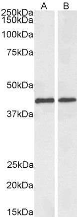
Figure 1. Western blot analysis of ACAT1 using anti-ACAT1 antibody (A02008-1). Electrophoresis was performed on a 5-20% SDS-PAGE gel at 70V (Stacking gel) / 90V (Resolving gel) for 2-3 hours. The sample well of each lane was loaded with 30 ug of sample under reducing conditions. Lane 1: human HepG2 whole cell lysates, Lane 2: human PC-3 whole cell lysates, Lane 3: human A549 whole cell lysates, Lane 4: human MCF-7 whole cell lysates, Lane 5: rat heart tissue lysates, Lane 6: rat skeletal muscle tissue lysates, Lane 7: mouse heart tissue lysates. After electrophoresis, proteins were transferred to a nitrocellulose membrane at 150 mA for 50-90 minutes. Blocked the membrane with 5% non-fat milk/TBS for 1.5 hour at RT. The membrane was incubated with rabbit anti-ACAT1 antigen affinity purified polyclonal antibody (Catalog # A02008-1) at 0.5 microg/mL overnight at 4°C, then washed with TBS-0.1%Tween 3 times with 5 minutes each and probed with a goat anti-rabbit IgG-HRP secondary antibody at a dilution of 1:5000 for 1.5 hour at RT. The signal is developed using an Enhanced Chemiluminescent detection (ECL) kit (Catalog # EK1002) with Tanon 5200 system. A specific band was detected for ACAT1 at approximately 40 kDa. The expected band size for ACAT1 is at 40 kDa.
. ACAT1 was detected in a paraffin-embedded section of human liver cancer tissue. Heat mediated antigen retrieval was performed in EDTA buffer (pH 8.0, epitope retrieval solution). The tissue section was blocked with 10% goat serum. The tissue section was then incubated with 2 microg/ml rabbit anti-ACAT1 Antibody (A02008-1) overnight at 4°C. Peroxidase Conjugated Goat Anti-rabbit IgG was used as secondary antibody and incubated for 30 minutes at 37°C. The tissue section was developed using HRP Conjugated Rabbit IgG Super Vision Assay Kit (Catalog # SV0002) with DAB as the chromogen.")
. ACAT1 was detected in a paraffin-embedded section of human non-small cell lung cancer tissue. Heat mediated antigen retrieval was performed in EDTA buffer (pH 8.0, epitope retrieval solution). The tissue section was blocked with 10% goat serum. The tissue section was then incubated with 2 microg/ml rabbit anti-ACAT1 Antibody (A02008-1) overnight at 4°C. Peroxidase Conjugated Goat Anti-rabbit IgG was used as secondary antibody and incubated for 30 minutes at 37°C. The tissue section was developed using HRP Conjugated Rabbit IgG Super Vision Assay Kit (Catalog # SV0002) with DAB as the chromogen.")
. ACAT1 was detected in a paraffin-embedded section of human placenta tissue. Heat mediated antigen retrieval was performed in EDTA buffer (pH 8.0, epitope retrieval solution). The tissue section was blocked with 10% goat serum. The tissue section was then incubated with 2 microg/ml rabbit anti-ACAT1 Antibody (A02008-1) overnight at 4°C. Peroxidase Conjugated Goat Anti-rabbit IgG was used as secondary antibody and incubated for 30 minutes at 37°C. The tissue section was developed using HRP Conjugated Rabbit IgG Super Vision Assay Kit (Catalog # SV0002) with DAB as the chromogen.")
. ACAT1 was detected in an immunocytochemical section of HELA cells. Enzyme antigen retrieval was performed using IHC enzyme antigen retrieval reagent (AR0022) for 15 mins. The cells were blocked with 10% goat serum. And then incubated with 5 microg/mL rabbit anti-ACAT1 Antibody (A02008-1) overnight at 4°C. Cy3 Conjugated Goat Anti-Rabbit IgG (BA1032) was used as secondary antibody at 1:500 dilution and incubated for 30 minutes at 37°C. The section was counterstained with DAPI. Visualize using a fluorescence microscope and filter sets appropriate for the label used.")
. Overlay histogram showing MCF-7 cells stained with A02008-1 (Blue line). To facilitate intracellular staining, cells were fixed with 4% paraformaldehyde and permeabilized with permeabilization buffer. The cells were blocked with 10% normal goat serum. And then incubated with rabbit anti-ACAT1 Antibody (A02008-1, 1 microg/1x106 cells) for 30 min at 20°C. DyLight®488 conjugated goat anti-rabbit IgG (BA1127, 5-10 microg/1x106 cells) was used as secondary antibody for 30 minutes at 20°C. Isotype control antibody (Green line) was rabbit IgG (1 microg/1x106) used under the same conditions. Unlabelled sample (Red line) was also used as a control.")
Figure 1. Western blot analysis of ACAT1 using anti-ACAT1 antibody (A02008-1). Electrophoresis was performed on a 5-20% SDS-PAGE gel at 70V (Stacking gel) / 90V (Resolving gel) for 2-3 hours. The sample well of each lane was loaded with 30 ug of sample under reducing conditions. Lane 1: human HepG2 whole cell lysates, Lane 2: human PC-3 whole cell lysates, Lane 3: human A549 whole cell lysates, Lane 4: human MCF-7 whole cell lysates, Lane 5: rat heart tissue lysates, Lane 6: rat skeletal muscle tissue lysates, Lane 7: mouse heart tissue lysates. After electrophoresis, proteins were transferred to a nitrocellulose membrane at 150 mA for 50-90 minutes. Blocked the membrane with 5% non-fat milk/TBS for 1.5 hour at RT. The membrane was incubated with rabbit anti-ACAT1 antigen affinity purified polyclonal antibody (Catalog # A02008-1) at 0.5 microg/mL overnight at 4°C, then washed with TBS-0.1%Tween 3 times with 5 minutes each and probed with a goat anti-rabbit IgG-HRP secondary antibody at a dilution of 1:5000 for 1.5 hour at RT. The signal is developed using an Enhanced Chemiluminescent detection (ECL) kit (Catalog # EK1002) with Tanon 5200 system. A specific band was detected for ACAT1 at approximately 40 kDa. The expected band size for ACAT1 is at 40 kDa.
Anti-ACAT1 Antibody Picoband(r)
A02008-1-CARRIER-FREE
ApplicationsFlow Cytometry, ImmunoFluorescence, Western Blot, ELISA, ImmunoCytoChemistry, ImmunoHistoChemistry
Product group Antibodies
ReactivityHuman, Mouse, Rat
TargetACAT1
Overview
- SupplierBoster Bio
- Product NameAnti-ACAT1 Antibody Picoband(r)
- Delivery Days Customer9
- ApplicationsFlow Cytometry, ImmunoFluorescence, Western Blot, ELISA, ImmunoCytoChemistry, ImmunoHistoChemistry
- CertificationResearch Use Only
- ClonalityPolyclonal
- Concentration500 ug/ml
- Gene ID38
- Target nameACAT1
- Target descriptionacetyl-CoA acetyltransferase 1
- Target synonymsACAT, MAT, T2, THIL, acetyl-CoA acetyltransferase, mitochondrial, acetoacetyl Coenzyme A thiolase, acetoacetyl-CoA thiolase, acetyl-Coenzyme A acetyltransferase 1, mitochondrial acetoacetyl-CoA thiolase, testicular tissue protein Li 198
- HostRabbit
- IsotypeIgG
- Protein IDP24752
- Protein NameAcetyl-CoA acetyltransferase, mitochondrial
- Scientific DescriptionBoster Bio Anti-ACAT1 Antibody Picoband® catalog # A02008-1. Tested in ELISA, IF, IHC, ICC, WB, Flow Cytometry applications. This antibody reacts with Human, Mouse, Rat. The brand Picoband indicates this is a premium antibody that guarantees superior quality, high affinity, and strong signals with minimal background in Western blot applications. Only our best-performing antibodies are designated as Picoband, ensuring unmatched performance.
- ReactivityHuman, Mouse, Rat
- Storage Instruction-20°C,2°C to 8°C
- UNSPSC12352203
Related products
Product group Antibodies
ApplicationsWestern Blot, ELISA
ReactivityHuman, Mouse, Rat
- SizePrice
Product group Antibodies
Anti-ACAT1 Antibody144-60258
ApplicationsWestern Blot, ImmunoHistoChemistry
ReactivityHuman, Mouse
TargetACAT1
- SizePrice
Product group Antibodies

ACAT1 AntibodyLS-C748331
ApplicationsWestern Blot, ImmunoHistoChemistry
ReactivityHuman, Mouse
TargetACAT1
- SizePrice
Product group Antibodies
ApplicationsFlow Cytometry, Western Blot, ImmunoCytoChemistry
ReactivityHuman, Rat
TargetACAT1
- SizePrice
Product group Antibodies
ACAT1 AntibodyCSB-PA001134ESR1HU
ApplicationsWestern Blot, ELISA, ImmunoHistoChemistry
ReactivityHuman, Mouse, Rat
TargetACAT1
- SizePrice
Product group Antibodies

ApplicationsImmunoFluorescence, Western Blot, ELISA
ReactivityBovine, Human, Mouse, Rat
TargetACAT1
- SizePrice
Product group Antibodies
ACAT1 Polyclonal AntibodyCAC14809
ApplicationsWestern Blot, ELISA, ImmunoHistoChemistry
ReactivityMouse, Rat
TargetACAT1
- SizePrice
Product group Antibodies
ACAT1 antibody [N1N3]GTX102637
ApplicationsImmunoFluorescence, Western Blot, ImmunoCytoChemistry, ImmunoHistoChemistry, ImmunoHistoChemistry Paraffin
ReactivityHuman, Mouse, Zebra Fish
TargetACAT1
- SizePrice

Product group Antibodies
Anti-ACAT1 AntibodyHPA004428
ApplicationsWestern Blot, ImmunoCytoChemistry, ImmunoHistoChemistry
ReactivityHuman, Mouse, Rat
TargetACAT1
- SizePrice