

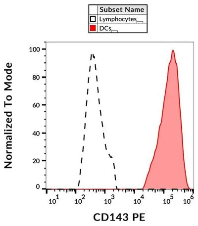
FACS analysis of human peripheral blood using GTX03649 ACE antibody [5-369] (PE).
FACS analysis of human peripheral blood using GTX03649 ACE antibody [5-369] (PE).
ACE antibody [5-369] (PE)
GTX03649
ApplicationsFlow Cytometry
Product group Antibodies
ReactivityHuman
TargetACE
Overview
- SupplierGeneTex
- Product NameACE antibody [5-369] (PE)
- Delivery Days Customer9
- Application Supplier NoteFCM: 10 microl reagent / 100 microl of whole blood or 106 cells in a suspension. *Optimal dilutions/concentrations should be determined by the researcher.Not tested in other applications.
- ApplicationsFlow Cytometry
- CertificationResearch Use Only
- ClonalityMonoclonal
- Clone ID5-369
- ConjugateRPE
- Gene ID1636
- Target nameACE
- Target descriptionangiotensin I converting enzyme
- Target synonymsACE1, CD143, DCP, DCP1, angiotensin-converting enzyme, CD143 antigen, angiotensin I converting enzyme (peptidyl-dipeptidase A) 1, carboxycathepsin, dipeptidyl carboxypeptidase 1, dipeptidyl carboxypeptidase I, kininase II, peptidase P
- HostMouse
- IsotypeIgG1
- Protein IDP12821
- Protein NameAngiotensin-converting enzyme
- Scientific DescriptionThis gene encodes an enzyme involved in catalyzing the conversion of angiotensin I into a physiologically active peptide angiotensin II. Angiotensin II is a potent vasopressor and aldosterone-stimulating peptide that controls blood pressure and fluid-electrolyte balance. This enzyme plays a key role in the renin-angiotensin system. Many studies have associated the presence or absence of a 287 bp Alu repeat element in this gene with the levels of circulating enzyme or cardiovascular pathophysiologies. Multiple alternatively spliced transcript variants encoding different isoforms have been identified, and two most abundant spliced variants encode the somatic form and the testicular form, respectively, that are equally active. [provided by RefSeq, May 2010]
- ReactivityHuman
- Storage Instruction-20°C or -80°C,2°C to 8°C
- UNSPSC41116161
Datasheet
Related products
Product group Antibodies
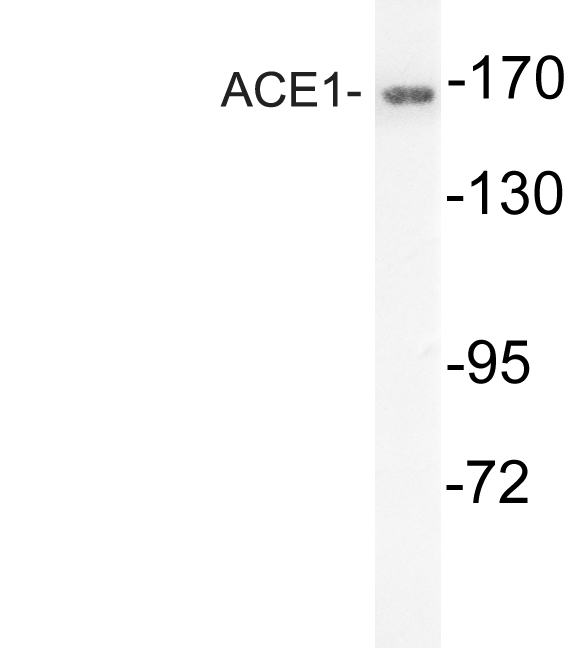
Anti-ACE1 AntibodyA96248
ApplicationsWestern Blot, ELISA, ImmunoHistoChemistry
ReactivityHuman, Mouse, Rat
- SizePrice
Product group Antibodies
Anti-ACE Antibody144-02805
ApplicationsWestern Blot
ReactivityHuman, Mouse
TargetACE
- SizePrice

Product group Antibodies
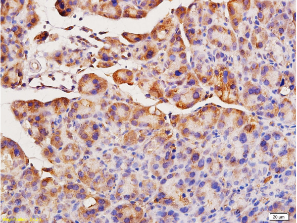
Anti-ACE Antibody Picoband(r)A00251-3-CARRIER-FREE
ApplicationsWestern Blot, ELISA, ImmunoHistoChemistry
ReactivityHuman, Mouse, Rat
TargetACE
- SizePrice
Product group Antibodies
References
ACE Polyclonal AntibodyBS-0439R
ApplicationsFlow Cytometry, ImmunoFluorescence, Western Blot, ELISA, ImmunoCytoChemistry, ImmunoHistoChemistry, ImmunoHistoChemistry Frozen, ImmunoHistoChemistry Paraffin
ReactivityBovine, Canine, Human, Mouse, Porcine, Rat
TargetACE
- SizePrice
Product group Antibodies
ACE AntibodyCSB-PA007661
ApplicationsWestern Blot, ELISA, ImmunoHistoChemistry
ReactivityHuman, Mouse, Rat
TargetACE
- SizePrice
Product group Antibodies
ApplicationsWestern Blot, ELISA, ImmunoCytoChemistry, ImmunoHistoChemistry, ImmunoHistoChemistry Frozen, ImmunoHistoChemistry Paraffin
TargetACE
- SizePrice

Product group Antibodies
Anti-ACE AntibodyHPA069790
ApplicationsImmunoCytoChemistry
ReactivityHuman
TargetACE
- SizePrice
Product group Antibodies
ACE antibodyGTX130534
ApplicationsWestern Blot, ImmunoHistoChemistry, ImmunoHistoChemistry Paraffin
ReactivityHuman, Mouse, Rat
TargetACE
- SizePrice
![WB analysis of human lung tissue using GTX52523 ACE antibody [12J16].](https://www.genetex.com/upload/website/prouct_img/normal/GTX52523/GTX52523_20191119_WB_w_23060900_186.webp)
Product group Antibodies
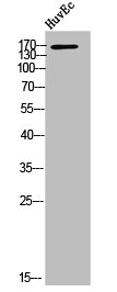
ACE antibody [12J16]GTX52523
ApplicationsWestern Blot
ReactivityHuman
TargetACE
- SizePrice
Product group Antibodies
ACE antibodyGTX54938
ApplicationsWestern Blot, ImmunoHistoChemistry, ImmunoHistoChemistry Paraffin
ReactivityHuman, Mouse, Rat
TargetACE
- SizePrice