

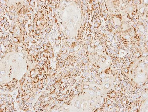
ACP2 antibody [N2C3] detects ACP2 protein at lysosome on Ca922 xenograft by immunohistochemical analysis. Sample: Paraffin-embedded Ca922 xenograft. ACP2 antibody [N2C3] (GTX114234) dilution: 1:500.
Antigen Retrieval: Trilogy? (EDTA based, pH 8.0) buffer, 15min
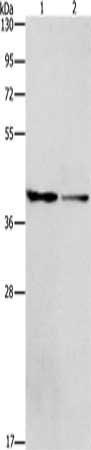
 A: A431 (GTX27909) 10% SDS PAGE Lysosomal acid phosphatase2 antibody GTX114234 diluted at 1:1000")
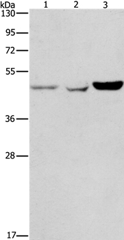
 antibody at 1:200 dilution.")
ACP2 antibody [N2C3] detects ACP2 protein at lysosome on Ca922 xenograft by immunohistochemical analysis. Sample: Paraffin-embedded Ca922 xenograft. ACP2 antibody [N2C3] (GTX114234) dilution: 1:500.
Antigen Retrieval: Trilogy? (EDTA based, pH 8.0) buffer, 15min
ACP2 antibody [N2C3]
GTX114234
ApplicationsImmunoFluorescence, Western Blot, ImmunoCytoChemistry, ImmunoHistoChemistry, ImmunoHistoChemistry Paraffin
Product group Antibodies
ReactivityHuman
TargetACP2
Overview
- SupplierGeneTex
- Product NameACP2 antibody [N2C3]
- Delivery Days Customer9
- Application Supplier NoteWB: 1:500-1:3000. ICC/IF: 1:100-1:1000. IHC-P: 1:100-1:1000. *Optimal dilutions/concentrations should be determined by the researcher.Not tested in other applications.
- ApplicationsImmunoFluorescence, Western Blot, ImmunoCytoChemistry, ImmunoHistoChemistry, ImmunoHistoChemistry Paraffin
- CertificationResearch Use Only
- ClonalityPolyclonal
- Concentration1 mg/ml
- ConjugateUnconjugated
- Gene ID53
- Target nameACP2
- Target descriptionacid phosphatase 2, lysosomal
- Target synonymsLAP, lysosomal acid phosphatase
- HostRabbit
- IsotypeIgG
- Protein IDP11117
- Protein NameLysosomal acid phosphatase
- Scientific DescriptionThis gene encodes the beta subunit of lysosomal acid phosphatase (LAP). LAP is chemically and genetically distinct from red cell acid phosphatase. The encoded protein belongs to a family of distinct isoenzymes which hydrolyze orthophosphoric monoesters to alcohol and phosphate. Mutations in this gene or in the related alpha subunit gene cause acid phosphatase deficiency. Multiple alternatively spliced transcript variants encoding different isoforms have been identified for this gene. [provided by RefSeq]
- ReactivityHuman
- Storage Instruction-20°C or -80°C,2°C to 8°C
- UNSPSC41116161
Datasheet
Related products
Product group Antibodies
ACP2 AntibodyCSB-PA240234
ApplicationsWestern Blot, ELISA
ReactivityHuman, Mouse, Rat
TargetACP2
- SizePrice

Product group Antibodies
Anti-ACP2 Antibody Picoband(r)A06554-1-CARRIER-FREE
ApplicationsWestern Blot
ReactivityHuman, Mouse, Rat
TargetACP2
- SizePrice
Product group Antibodies

ACP2 / Acid Phosphatase 2 AntibodyLS-C659388
ApplicationsWestern Blot, ELISA
ReactivityHuman
TargetACP2
- SizePrice
Product group Antibodies
ApplicationsImmunoPrecipitation, Western Blot, ImmunoCytoChemistry, ImmunoHistoChemistry
ReactivityMouse, Rat
TargetACP2
- SizePrice
Product group Antibodies
Anti-ACP2 Antibody101-10017
ApplicationsImmunoFluorescence, Western Blot, ELISA, ImmunoHistoChemistry
TargetACP2
- SizePrice