

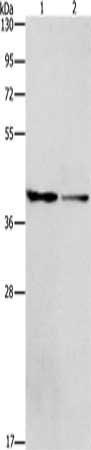
Anti-ACP2 Antibody
101-10017
ApplicationsImmunoFluorescence, Western Blot, ELISA, ImmunoHistoChemistry
Product group Antibodies
TargetACP2
Overview
- SupplierRayBiotech
- Product NameAnti-ACP2 Antibody
- Delivery Days Customer4
- ApplicationsImmunoFluorescence, Western Blot, ELISA, ImmunoHistoChemistry
- CertificationResearch Use Only
- ClonalityMonoclonal
- Gene ID53
- Target nameACP2
- Target descriptionacid phosphatase 2, lysosomal
- Target synonymsLAP, lysosomal acid phosphatase
- HostMouse
- IsotypeIgG2b
- Scientific DescriptionHuman ACP2 monoclonal antibody (100 ug)
- Storage Instruction-20°C
- UNSPSC12352203
Related products
Product group Antibodies
ACP2 AntibodyCSB-PA240234
ApplicationsWestern Blot, ELISA
ReactivityHuman, Mouse, Rat
TargetACP2
- SizePrice

Product group Antibodies
Anti-ACP2 Antibody Picoband(r)A06554-1-CARRIER-FREE
ApplicationsWestern Blot
ReactivityHuman, Mouse, Rat
TargetACP2
- SizePrice
Product group Antibodies

ACP2 / Acid Phosphatase 2 AntibodyLS-C659388
ApplicationsWestern Blot, ELISA
ReactivityHuman
TargetACP2
- SizePrice
Product group Antibodies
ApplicationsImmunoPrecipitation, Western Blot, ImmunoCytoChemistry, ImmunoHistoChemistry
ReactivityMouse, Rat
TargetACP2
- SizePrice
![ACP2 antibody [N2C3] detects ACP2 protein at lysosome on Ca922 xenograft by immunohistochemical analysis. Sample: Paraffin-embedded Ca922 xenograft. ACP2 antibody [N2C3] (GTX114234) dilution: 1:500.
Antigen Retrieval: Trilogy? (EDTA based, pH 8.0) buffer, 15min](https://www.genetex.com/upload/website/prouct_img/normal/GTX114234/GTX114234_IHC_w_23060501_947.webp)
Product group Antibodies
ACP2 antibody [N2C3]GTX114234
ApplicationsImmunoFluorescence, Western Blot, ImmunoCytoChemistry, ImmunoHistoChemistry, ImmunoHistoChemistry Paraffin
ReactivityHuman
TargetACP2
- SizePrice