

Mouse heart lysates probed with Actin/ACTA1 Polyclonal Antibody, Unconjugated (bs-10966R) at 1:300 dilution and 4˚C overnight incubation. Followed by conjugated secondary antibody incubation at 1:20000 for 60 min at 37˚C.
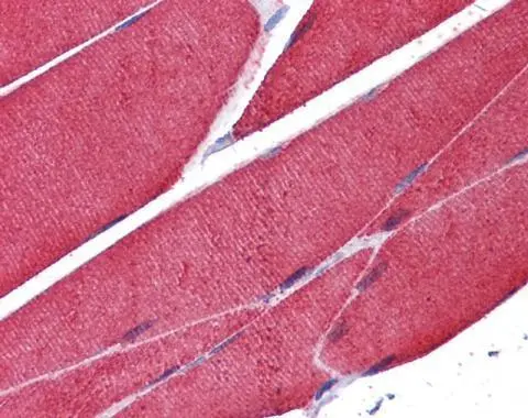
; Antigen retrieval by boiling in sodium citrate buffer (pH6.0) for 15min; Block endogenous peroxidase by 3% hydrogen peroxide for 20 minutes; Blocking buffer (normal goat serum) at 37°C for 30min; Antibody incubation with (Actin) Polyclonal Antibody, Unconjugated (bs-10966R) at 1:200 overnight at 4°C, followed by operating according to SP Kit(Rabbit) (sp-0023) instructionsand DAB staining.")
; Antigen retrieval by boiling in sodium citrate buffer (pH6.0) for 15min; Block endogenous peroxidase by 3% hydrogen peroxide for 20 minutes; Blocking buffer (normal goat serum) at 37°C for 30min; Antibody incubation with (Actin) Polyclonal Antibody, Unconjugated (bs-10966R) at 1:200 overnight at 4°C, followed by operating according to SP Kit(Rabbit) (sp-0023) instructionsand DAB staining.")
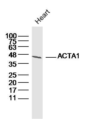
Mouse heart lysates probed with Actin/ACTA1 Polyclonal Antibody, Unconjugated (bs-10966R) at 1:300 dilution and 4˚C overnight incubation. Followed by conjugated secondary antibody incubation at 1:20000 for 60 min at 37˚C.
Actin Polyclonal Antibody
BS-10966R
ApplicationsImmunoFluorescence, Western Blot, ELISA, ImmunoCytoChemistry, ImmunoHistoChemistry, ImmunoHistoChemistry Frozen, ImmunoHistoChemistry Paraffin
Product group Antibodies
ReactivityBovine, Canine, Human, Mouse, Rabbit, Rat
TargetACTA1
Overview
- SupplierBioss
- Product NameActin Polyclonal Antibody
- Delivery Days Customer16
- ApplicationsImmunoFluorescence, Western Blot, ELISA, ImmunoCytoChemistry, ImmunoHistoChemistry, ImmunoHistoChemistry Frozen, ImmunoHistoChemistry Paraffin
- Applications SupplierWB(1:300-5000), ELISA(1:500-1000), IHC-P(1:200-400), IHC-F(1:100-500), IF(IHC-P)(1:50-200), IF(IHC-F)(1:50-200), IF(ICC)(1:50-200)
- CertificationResearch Use Only
- ClonalityPolyclonal
- Concentration1 ug/ul
- ConjugateUnconjugated
- Gene ID58
- Target nameACTA1
- Target descriptionactin alpha 1, skeletal muscle
- Target synonymsACTA, ASMA, CFTD, CFTD1, CFTDM, CMYO2A, CMYO2B, CMYO2C, CMYP2A, CMYP2B, CMYP2C, MPFD, NEM1, NEM2, NEM3, SHPM, actin, alpha skeletal muscle, nemaline myopathy type 3
- HostRabbit
- IsotypeIgG
- Protein IDP68133
- Protein NameActin, alpha skeletal muscle
- ReactivityBovine, Canine, Human, Mouse, Rabbit, Rat
- Storage Instruction-20°C
- UNSPSC41116161
References
- 3-Bromopyruvate inhibits the malignant phenotype of malignantly transformed macrophages and dendritic cells induced by glioma stem cells in the glioma microenvironment via miR-449a/MCT1. Sheng Y et al., 2020 Jan, Biomed PharmacotherRead this paper
- Chlorogenic acid attenuates cadmium-induced intestinal injury in Sprague-Dawley rats. Xue Y et al., 2019 Nov, Food Chem ToxicolRead this paper
Datasheet
Related products
Product group Antibodies

Anti-Alpha actin 1 [3G8C7A6]Ab03147-10.0
ApplicationsELISA, ImmunoHistoChemistry, ImmunoHistoChemistry Paraffin
ReactivityHuman
TargetACTA1
- SizePrice
Product group Antibodies
Anti-actin al Antibody130-10255
ApplicationsWestern Blot, ELISA
ReactivityHuman
TargetACTA1
- SizePrice
Product group Antibodies
ACTA1 Monoclonal AntibodyCSB-MA000309
ApplicationsImmunoPrecipitation, Western Blot, ELISA
ReactivityHuman, Mouse, Rat
TargetACTA1
- SizePrice
Product group Antibodies
Acta1 Polyclonal AntibodyCAC07080
ApplicationsWestern Blot, ELISA, ImmunoHistoChemistry
ReactivityMouse, Rat
TargetACTA1
- SizePrice
Product group Antibodies

Mouse anti actin alpha-skeletalMUB0108P
ApplicationsWestern Blot, ELISA, ImmunoHistoChemistry, ImmunoHistoChemistry Frozen, ImmunoHistoChemistry Paraffin
ReactivityHuman, Mammals, Porcine, Rabbit, Rat
TargetACTA1
- SizePrice
Product group Antibodies
alpha Skeletal Muscle Actin antibodyGTX101362
ApplicationsFlow Cytometry, Western Blot, ImmunoHistoChemistry, ImmunoHistoChemistry Paraffin
ReactivityHuman, Mouse, Rat
TargetACTA1
- SizePrice
Product group Antibodies

ApplicationsWestern Blot, ELISA
ReactivityHuman
TargetACTA1
- SizePrice

Product group Antibodies
Anti-Actin/ACTA1 Antibody Picoband(r)PB9751-CARRIER-FREE
ApplicationsWestern Blot, ImmunoHistoChemistry
ReactivityHamster, Human, Mouse, Rat
TargetACTA1
- SizePrice
Product group Antibodies
TargetACTA1
- SizePrice