

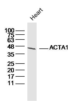
Mouse anti actin alpha-skeletal
MUB0108P
ApplicationsWestern Blot, ELISA, ImmunoHistoChemistry, ImmunoHistoChemistry Frozen, ImmunoHistoChemistry Paraffin
Product group Antibodies
ReactivityHuman, Mammals, Porcine, Rabbit, Rat
TargetACTA1
Overview
- SupplierNordic-MUbio
- Product NameMouse anti actin alpha-skeletal
- Delivery Days Customer7
- Application Supplier Note3B3 is useful for immunohistochemistry on frozen and paraffin-embedded tissues, immunoblotting and ELISA. Optimal antibody dilutions should be determined by titration; recommended range is 1:100 - 1:1000 for immunohistochemistry with avidin-biotinylated Horseradish peroxidase complex (ABC) as detection reagent, and 1:1000 - 1:5000 for immunoblotting applications.
- ApplicationsWestern Blot, ELISA, ImmunoHistoChemistry, ImmunoHistoChemistry Frozen, ImmunoHistoChemistry Paraffin
- Applications SupplierELISA;Immunohistochemistry (frozen);Immunohistochemistry (paraffin);Western Blotting
- CertificationResearch Use Only
- ClonalityMonoclonal
- Clone ID3B3
- Gene ID58
- Target nameACTA1
- Target descriptionactin alpha 1, skeletal muscle
- Target synonymsACTA, ASMA, CFTD, CFTD1, CFTDM, CMYO2A, CMYO2B, CMYO2C, CMYP2A, CMYP2B, CMYP2C, MPFD, NEM1, NEM2, NEM3, SHPM, actin, alpha skeletal muscle, nemaline myopathy type 3
- HostMouse
- IsotypeIgG1
- Protein IDP68133
- Protein NameActin, alpha skeletal muscle
- Source3B3 is a Mouse monoclonal IgG1 antibody derived by fusion of NS0 Mouse myeloma cells with spleen cells from a BALB/c Mouse immunized with a peptide comprising the N-terminal nonapeptide of alpha-skeletal actin with an acetylated N-terminus ...
- ReactivityHuman, Mammals, Porcine, Rabbit, Rat
- Reactivity SupplierCaprine;Human;Rabbit;Rat;Swine
- UNSPSC12352203
Related products
Product group Antibodies

Anti-Alpha actin 1 [3G8C7A6]Ab03147-10.0
ApplicationsELISA, ImmunoHistoChemistry, ImmunoHistoChemistry Paraffin
ReactivityHuman
TargetACTA1
- SizePrice
Product group Antibodies
Anti-actin al Antibody130-10255
ApplicationsWestern Blot, ELISA
ReactivityHuman
TargetACTA1
- SizePrice
Product group Antibodies
References
Actin Polyclonal AntibodyBS-10966R
ApplicationsImmunoFluorescence, Western Blot, ELISA, ImmunoCytoChemistry, ImmunoHistoChemistry, ImmunoHistoChemistry Frozen, ImmunoHistoChemistry Paraffin
ReactivityBovine, Canine, Human, Mouse, Rabbit, Rat
TargetACTA1
- SizePrice
Product group Antibodies
ACTA1 Monoclonal AntibodyCSB-MA000309
ApplicationsImmunoPrecipitation, Western Blot, ELISA
ReactivityHuman, Mouse, Rat
TargetACTA1
- SizePrice
Product group Antibodies
Acta1 Polyclonal AntibodyCAC07080
ApplicationsWestern Blot, ELISA, ImmunoHistoChemistry
ReactivityMouse, Rat
TargetACTA1
- SizePrice

Product group Antibodies
alpha Skeletal Muscle Actin antibodyGTX101362
ApplicationsFlow Cytometry, Western Blot, ImmunoHistoChemistry, ImmunoHistoChemistry Paraffin
ReactivityHuman, Mouse, Rat
TargetACTA1
- SizePrice
Product group Antibodies

ApplicationsWestern Blot, ELISA
ReactivityHuman
TargetACTA1
- SizePrice

Product group Antibodies
Anti-Actin/ACTA1 Antibody Picoband(r)PB9751-CARRIER-FREE
ApplicationsWestern Blot, ImmunoHistoChemistry
ReactivityHamster, Human, Mouse, Rat
TargetACTA1
- SizePrice
Product group Antibodies
TargetACTA1
- SizePrice