

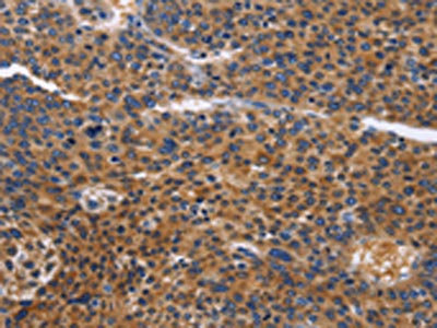
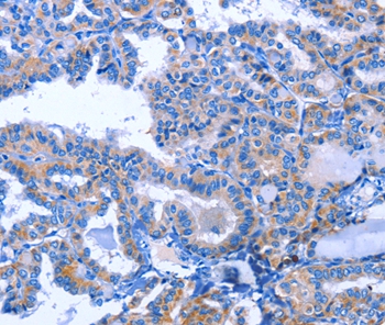
The image on the left is immunohistochemistry of paraffin-embedded Human liver cancer tissue using CSB-PA206692(ACVR2B Antibody) at dilution 1/50, on the right is treated with fusion protein. (Original magnification: x200)
 at dilution 1/50, on the right is treated with fusion protein. (Original magnification: x200)")
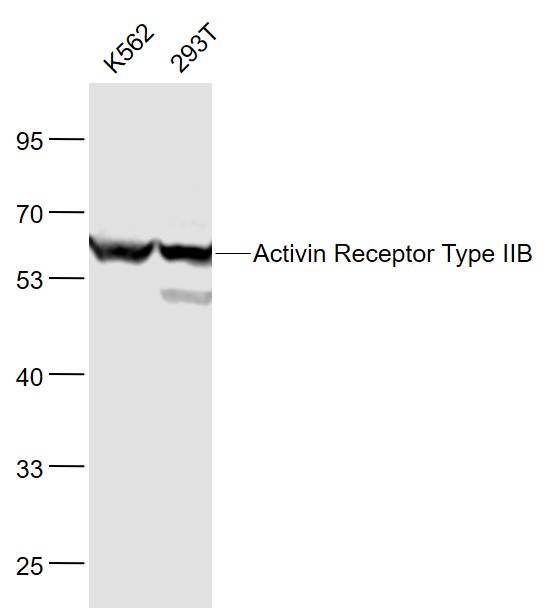
 at dilution 1/300, Secondary antibody: Goat anti rabbit IgG at 1/8000 dilution, Exposure time: 20 seconds")
The image on the left is immunohistochemistry of paraffin-embedded Human liver cancer tissue using CSB-PA206692(ACVR2B Antibody) at dilution 1/50, on the right is treated with fusion protein. (Original magnification: x200)
ACVR2B Antibody
CSB-PA206692
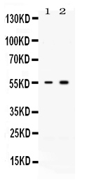
ApplicationsWestern Blot, ELISA, ImmunoHistoChemistry
Product group Antibodies
ReactivityHuman, Mouse, Rat
TargetACVR2B
Overview
- SupplierCusabio
- Product NameACVR2B Antibody
- Delivery Days Customer20
- ApplicationsWestern Blot, ELISA, ImmunoHistoChemistry
- CertificationResearch Use Only
- ClonalityPolyclonal
- ConjugateUnconjugated
- Gene ID93
- Target nameACVR2B
- Target descriptionactivin A receptor type 2B
- Target synonymsACTRIIB, ActR-IIB, HTX4, activin receptor type-2B, activin A receptor, type IIB
- HostRabbit
- IsotypeIgG
- Protein IDQ13705
- Protein NameActivin receptor type-2B
- Scientific DescriptionActivins are dimeric growth and differentiation factors which belong to the transforming growth factor-beta (TGF-beta) superfamily of structurally related signaling proteins. Activins signal through a heteromeric complex of receptor serine kinases which include at least two type I (I and IB) and two type II (II and IIB) receptors. These receptors are all transmembrane proteins, composed of a ligand-binding extracellular domain with cysteine-rich region, a transmembrane domain, and a cytoplasmic domain with predicted serine/threonine specificity. Type I receptors are essential for signaling; and type II receptors are required for binding ligands and for expression of type I receptors. Type I and II receptors form a stable complex after ligand binding, resulting in phosphorylation of type I receptors by type II receptors. Type II receptors are considered to be constitutively active kinases. This gene encodes activin A type IIB receptor, which displays a 3- to 4-fold higher affinity for the ligand than activin A type II receptor.
- ReactivityHuman, Mouse, Rat
- Storage Instruction-20°C or -80°C
- UNSPSC41116161
Related products
Product group Antibodies
Anti-ACVR2B AntibodyA45381
ApplicationsImmunoHistoChemistry
ReactivityHuman
- SizePrice
Product group Antibodies
Anti-ACVR2B Antibody144-66500
ApplicationsImmunoFluorescence, Western Blot
ReactivityHuman
TargetACVR2B
- SizePrice
Product group Antibodies
ApplicationsImmunoFluorescence, Western Blot, ELISA, ImmunoCytoChemistry, ImmunoHistoChemistry, ImmunoHistoChemistry Frozen, ImmunoHistoChemistry Paraffin
ReactivityBovine, Canine, Human, Mouse, Porcine, Rat, Sheep
TargetACVR2B
- SizePrice
Product group Antibodies
Acvr2B Recombinant AntibodyCAC11965
ApplicationsELISA, ImmunoHistoChemistry
TargetACVR2B
- SizePrice
Product group Antibodies

ACTRIIB / ACVR2B AntibodyLS-C483187
ApplicationsELISA
ReactivityMouse
TargetACVR2B
- SizePrice
![Activin Receptor Type IIB antibody [N1C1] detects Activin Receptor Type IIB protein at cytoplasm by immunohistochemical analysis. Sample: Paraffin-embedded mouse placenta. Activin Receptor Type IIB stained by Activin Receptor Type IIB antibody [N1C1] (GTX105330) diluted at 1:500. Antigen Retrieval: Citrate buffer, pH 6.0, 15 min](https://www.genetex.com/upload/website/prouct_img/normal/GTX105330/GTX105330_43404_20190118_IHC-P_M_w_23060120_824.webp)
Product group Antibodies
ApplicationsWestern Blot, ImmunoHistoChemistry, ImmunoHistoChemistry Paraffin
ReactivityHuman, Mouse
TargetACVR2B
- SizePrice
Product group Antibodies
TargetACVR2B
- SizePrice
Product group Antibodies
Anti-Activin Receptor Type IIB/ACVR2B Antibody Picoband(r)PB9975-CARRIER-FREE
ApplicationsWestern Blot
ReactivityBovine, Human, Rat
TargetACVR2B
- SizePrice
Product group Antibodies
Anti-ACVR2B AntibodyCAB7868
ApplicationsImmunoFluorescence, Western Blot, ELISA, ImmunoCytoChemistry
ReactivityHuman
TargetACVR2B
- SizePrice