

Anti-ADAR1 Antibody
CAB7869
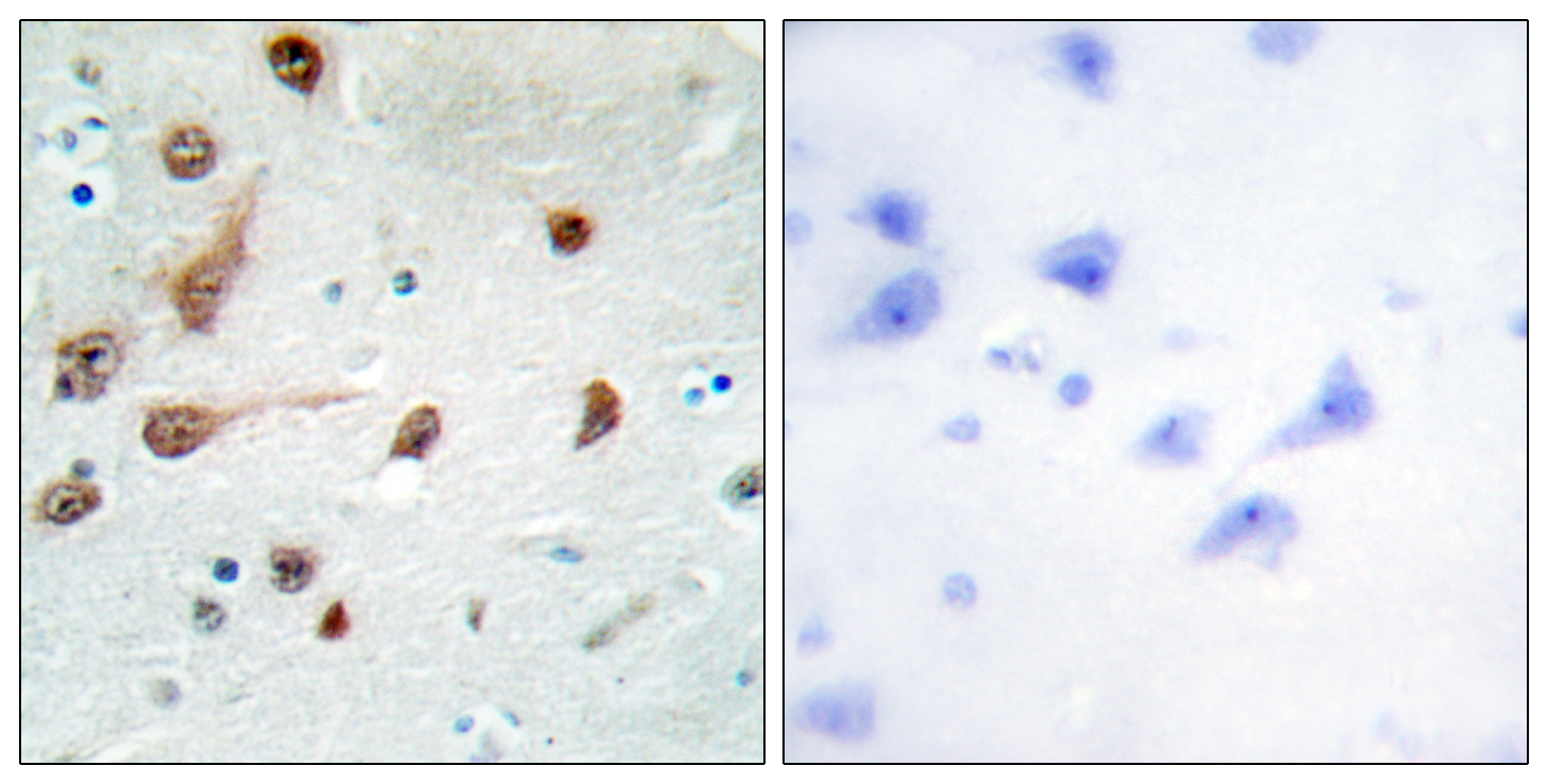
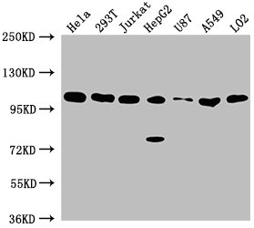
ApplicationsImmunoFluorescence, Western Blot, ELISA, ImmunoCytoChemistry, ImmunoHistoChemistry, ImmunoHistoChemistry Paraffin
Product group Antibodies
ReactivityHuman
TargetADAR
Overview
- SupplierAssay Genie
- Product NameAnti-ADAR1 Antibody
- Delivery Days Customer9
- ApplicationsImmunoFluorescence, Western Blot, ELISA, ImmunoCytoChemistry, ImmunoHistoChemistry, ImmunoHistoChemistry Paraffin
- Applications SupplierWB,IHC
- CertificationResearch Use Only
- ClonalityPolyclonal
- Clone IDNot applicable
- ConjugateUnconjugated
- Gene ID103
- Target nameADAR
- Target descriptionadenosine deaminase RNA specific
- Target synonymsADAR1, AGS6, DRADA, DSH, DSRAD, G1P1, IFI-4, IFI4, K88DSRBP, P136, double-stranded RNA-specific adenosine deaminase, 136 kDa double-stranded RNA-binding protein, adenosine deaminase acting on RNA 1-A, dsRNA adenosine deaminase, dsRNA adeonosine deaminase, interferon-induced protein 4, interferon-inducible protein 4
- HostRabbit
- Protein IDP55265
- Protein NameDouble-stranded RNA-specific adenosine deaminase
- Scientific DescriptionADAR1 Antibody is a premium polyclonal antibody that offers outstanding performance and reliability for demanding research applications. Rigorously validated for WB, IHC-P, IF/ICC, ELISA, this antibody ensures consistent, reproducible results across multiple experimental platforms. Demonstrates excellent reactivity with Human,Mouse,Rat samples, providing researchers with confidence in cross-species compatibility. Conveniently packaged in 100microL format to meet your experimental needs. For optimal performance, store at -20°C and maintains stability for 12 months. Backed by rigorous quality control testing to ensure superior performance in your critical research applications.
- Shelf life instruction12 months
- SourceRabbit
- ReactivityHuman
- Reactivity SupplierHuman,Mouse,Rat
- Storage Instruction-20°C
- UNSPSC41116161
Related products
Product group Antibodies
Anti-ADAR1 AntibodyA97053
ApplicationsELISA, ImmunoHistoChemistry
ReactivityHuman, Mouse, Rat
- SizePrice

Product group Antibodies
Anti-ADAR1/ADAR Antibody Picoband(r)A00869-2-CARRIER-FREE
ApplicationsImmunoFluorescence, Western Blot, ELISA, ImmunoCytoChemistry, ImmunoHistoChemistry
ReactivityHuman, Mouse, Rat
TargetADAR
- SizePrice
Product group Antibodies
Anti-ADAR Antibody144-07869
ApplicationsImmunoFluorescence, Western Blot, ImmunoHistoChemistry
ReactivityHuman, Mouse, Rat
TargetADAR
- SizePrice

Product group Antibodies
Anti-ADAR AntibodyAMAB90535
ApplicationsWestern Blot, ImmunoCytoChemistry, ImmunoHistoChemistry
ReactivityHuman
TargetADAR
- SizePrice
Product group Antibodies
ADAR1 Polyclonal AntibodyBS-2168R
ApplicationsImmunoFluorescence, Western Blot, ELISA, ImmunoCytoChemistry, ImmunoHistoChemistry, ImmunoHistoChemistry Frozen, ImmunoHistoChemistry Paraffin
ReactivityHuman, Mouse, Rat
TargetADAR
- SizePrice
Product group Antibodies
ADAR AntibodyCSB-PA001324LA01HU
ApplicationsWestern Blot, ELISA, ImmunoHistoChemistry
ReactivityHuman
TargetADAR
- SizePrice
Product group Antibodies
Adar Polyclonal AntibodyCAC11643
ApplicationsWestern Blot, ELISA, ImmunoHistoChemistry
TargetADAR
- SizePrice
![ADAR1 antibody [N3C1], Internal detects ADAR1 protein at nucleolus and nucleus by immunofluorescent analysis. Sample: HeLa cells were fixed in 4% paraformaldehyde at RT for 15 min. Green: ADAR1 protein stained by ADAR1 antibody [N3C1], Internal (GTX101602) diluted at 1:200. Red: phalloidin, a cytoskeleton marker, diluted at 1:200. Blue: Hoechst 33342 staining. Scale bar = 10 μm.](https://www.genetex.com/upload/website/prouct_img/normal/GTX101602/GTX101602_40506_20150714_IFA_w_23060100_395.webp)
Product group Antibodies
ADAR1 antibody [N3C1], InternalGTX101602
ApplicationsImmunoFluorescence, ImmunoPrecipitation, Western Blot, ImmunoCytoChemistry, ImmunoHistoChemistry, ImmunoHistoChemistry Paraffin
ReactivityHuman, Rat
TargetADAR
- SizePrice