

AFAP Polyclonal Antibody
BS-9790R
ApplicationsImmunoFluorescence, Western Blot, ImmunoHistoChemistry, ImmunoHistoChemistry Paraffin
Product group Antibodies
ReactivityBovine, Canine, Equine, Human, Mouse, Rat, Sheep
TargetAFAP1
Overview
- SupplierBioss
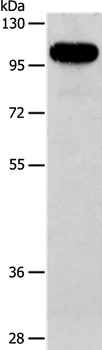
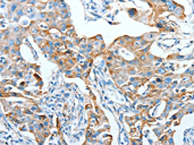
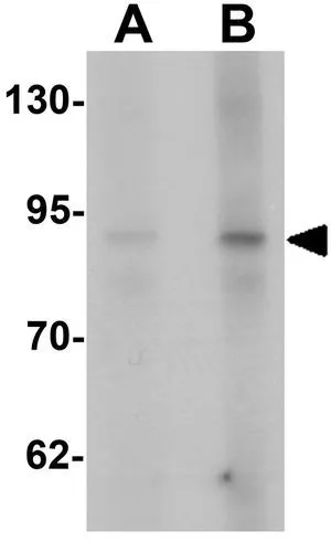
- Product NameAFAP Polyclonal Antibody
- Delivery Days Customer16
- ApplicationsImmunoFluorescence, Western Blot, ImmunoHistoChemistry, ImmunoHistoChemistry Paraffin
- Applications SupplierWB(1:300-5000), IHC-P(1:200-400), IF(IHC-P)(1:50-200)
- CertificationResearch Use Only
- ClonalityPolyclonal
- Concentration1 ug/ul
- ConjugateUnconjugated
- Gene ID60312
- Target nameAFAP1
- Target descriptionactin filament associated protein 1
- Target synonymsAFAP, AFAP-110, AFAP110, actin filament-associated protein 1, 110 kDa actin filament-associated protein, actin filament-associated protein, 110 kDa
- HostRabbit
- IsotypeIgG
- Protein IDQ8N556
- Protein NameActin filament-associated protein 1
- ReactivityBovine, Canine, Equine, Human, Mouse, Rat, Sheep
- Storage Instruction-20°C
- UNSPSC41116161
Datasheet
Related products
Product group Antibodies
Anti-AFAP1 AntibodyA37858
ApplicationsWestern Blot, ImmunoHistoChemistry
ReactivityHuman
- SizePrice

Product group Antibodies
Anti-AFAP/AFAP1 Antibody Picoband(r)A05258-3-CARRIER-FREE
ApplicationsFlow Cytometry, Western Blot, ELISA, ImmunoHistoChemistry
ReactivityHuman, Mouse, Rat
TargetAFAP1
- SizePrice
Product group Antibodies
Anti-AFAP1 Antibody144-03478
ApplicationsWestern Blot
ReactivityHuman, Mouse, Rat
TargetAFAP1
- SizePrice
Product group Antibodies
AFAP1 AntibodyCSB-PA098902
ApplicationsWestern Blot, ELISA, ImmunoHistoChemistry
ReactivityHuman, Mouse, Rat
TargetAFAP1
- SizePrice
Product group Antibodies
AFAP antibodyGTX31462
ApplicationsWestern Blot, ELISA
ReactivityHuman, Mouse
TargetAFAP1
- SizePrice
Product group Antibodies

AFAP1 / AFAP AntibodyLS-C332595
ApplicationsWestern Blot
ReactivityHuman, Mouse, Rat
TargetAFAP1
- SizePrice

Product group Antibodies
Anti-AFAP1 AntibodyHPA015642
ApplicationsImmunoCytoChemistry, ImmunoHistoChemistry
ReactivityHuman
TargetAFAP1
- SizePrice
Product group Antibodies
Anti-AFAP1 AntibodyCAB3478
ApplicationsImmunoFluorescence, Western Blot, ELISA, ImmunoCytoChemistry
ReactivityHuman
TargetAFAP1
- SizePrice