

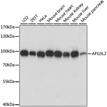
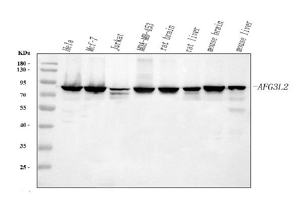
WB analysis of various sample lysates using GTX66373 AFG3L2 antibody. Dilution : 1:1000 Loading : 25μg per lane


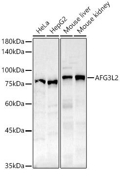
WB analysis of various sample lysates using GTX66373 AFG3L2 antibody. Dilution : 1:1000 Loading : 25μg per lane
AFG3L2 antibody
GTX66373
ApplicationsWestern Blot, ImmunoHistoChemistry, ImmunoHistoChemistry Paraffin
Product group Antibodies
ReactivityHuman, Mouse, Rat
TargetAFG3L2
Overview
- SupplierGeneTex
- Product NameAFG3L2 antibody
- Delivery Days Customer9
- Application Supplier NoteWB: 1:200 - 1:2000. IHC-P: 1:50 - 1:200. *Optimal dilutions/concentrations should be determined by the researcher.Not tested in other applications.
- ApplicationsWestern Blot, ImmunoHistoChemistry, ImmunoHistoChemistry Paraffin
- CertificationResearch Use Only
- ClonalityPolyclonal
- ConjugateUnconjugated
- Gene ID10939
- Target nameAFG3L2
- Target descriptionAFG3 like matrix AAA peptidase subunit 2
- Target synonymsOPA12, SCA28, SPAX5, mitochondrial inner membrane m-AAA protease component AFG3L2, AFG3 ATPase family gene 3-like 2, AFG3 ATPase family member 3-like 2, AFG3 like AAA ATPase 2, AFG3-like protein 2, ATPase family gene 3, yeast, paraplegin-like protein
- HostRabbit
- IsotypeIgG
- Protein IDQ9Y4W6
- Protein NameMitochondrial inner membrane m-AAA protease component AFG3L2
- Scientific DescriptionThis gene encodes a protein localized in mitochondria and closely related to paraplegin. The paraplegin gene is responsible for an autosomal recessive form of hereditary spastic paraplegia. This gene is a candidate gene for other hereditary spastic paraplegias or neurodegenerative disorders. [provided by RefSeq, Jul 2008]
- ReactivityHuman, Mouse, Rat
- Storage Instruction-20°C or -80°C,2°C to 8°C
- UNSPSC41116161
Datasheet
Related products
Product group Antibodies
Anti-AFG3L2 AntibodyA91394
ApplicationsImmunoPrecipitation, Western Blot, ImmunoHistoChemistry
ReactivityHuman, Mouse, Rat
- SizePrice
Product group Antibodies
Anti-AFG3L2 Antibody144-61032
ApplicationsWestern Blot, ImmunoHistoChemistry
ReactivityHuman, Mouse, Rat
TargetAFG3L2
- SizePrice
Product group Antibodies

AFG3L2 AntibodyLS-C750363
ApplicationsWestern Blot
ReactivityHuman, Mouse
TargetAFG3L2
- SizePrice
Product group Antibodies
AFG3L2 Polyclonal AntibodyCAC14055
ApplicationsWestern Blot, ELISA, ImmunoHistoChemistry
TargetAFG3L2
- SizePrice
Product group Antibodies
AFG3L2 AntibodyCSB-PA04005A0RB
ApplicationsWestern Blot, ELISA, ImmunoHistoChemistry
ReactivityHuman
TargetAFG3L2
- SizePrice
Product group Antibodies
AFG3L2 antibody [N1N2], N-termGTX102036
ApplicationsWestern Blot, ImmunoHistoChemistry, ImmunoHistoChemistry Paraffin
ReactivityHuman, Rat
TargetAFG3L2
- SizePrice

Product group Antibodies
Anti-AFG3L2 AntibodyHPA004479
ApplicationsImmunoCytoChemistry, ImmunoHistoChemistry
ReactivityHuman
TargetAFG3L2
- SizePrice
Product group Antibodies
Anti-AFG3L2 Antibody Picoband(r)PB9915-CARRIER-FREE
ApplicationsFlow Cytometry, ImmunoFluorescence, ImmunoPrecipitation, Western Blot, ImmunoCytoChemistry
ReactivityHuman, Mouse, Rat
TargetAFG3L2
- SizePrice