

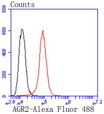
Flow cytometric analysis of HeLa cells with AGR2 (3F8) Monoclonal Antibody (bsm-52594R) at 1:50 dilution (red) compared with an unlabeled control (cells without incubation with primary antibody; black).
 Monoclonal Antibody (bsm-52594R) at 1:1000 overnight at 4°C followed by a conjugated secondary antibody for 60 minutes at 37°C.")
 staining with AGR2 (3F8) Monoclonal Antibody (bsm-52594R) at 1:300 in HeLa cells (green). The nuclear counterstain is DAPI (blue). Cells were fixed in paraformaldehyde, permeabilized with 0.25% Triton X100/PBS.")
 staining with AGR2 (3F8) Monoclonal Antibody (bsm-52594R) at 1:300 in MCF-7 cells (green). The nuclear counterstain is DAPI (blue). Cells were fixed in paraformaldehyde, permeabilized with 0.25% Triton X100/PBS.")
 staining with AGR2 (3F8) Monoclonal Antibody (bsm-52594R) at 1:300 in NIH/3T3 cells (green). The nuclear counterstain is DAPI (blue). Cells were fixed in paraformaldehyde, permeabilized with 0.25% Triton X100/PBS.")
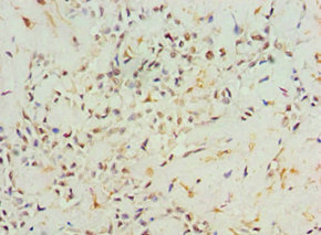
 Monoclonal Antibody (bsm-52594R) at 1:100, overnight at 4°C, followed by a conjugated secondary antibody and DAB staining. Counterstained with hematoxylin.")
 for 15min; Block endogenous peroxidase by 3% hydrogen peroxide for 20 minutes; Blocking buffer (normal goat serum) at 37°C for 30min; Antibody incubation with AGR2 (3F8) Monoclonal Antibody, Unconjugated (bsm-52594R) at 1:200 overnight at 4°C, DAB staining.")
 for 15min; Block endogenous peroxidase by 3% hydrogen peroxide for 20 minutes; Blocking buffer (normal goat serum) at 37°C for 30min; Antibody incubation with AGR2 (3F8) Monoclonal Antibody, Unconjugated (bsm-52594R) at 1:200 overnight at 4°C, DAB staining.")
 for 15min; Block endogenous peroxidase by 3% hydrogen peroxide for 20 minutes; Blocking buffer (normal goat serum) at 37°C for 30min; Antibody incubation with AGR2 (3F8) Monoclonal Antibody, Unconjugated (bsm-52594R) at 1:200 overnight at 4°C, DAB staining.")
 for 15min; Block endogenous peroxidase by 3% hydrogen peroxide for 20 minutes; Blocking buffer (normal goat serum) at 37°C for 30min; Antibody incubation with AGR2 (3F8) Monoclonal Antibody, Unconjugated (bsm-52594R) at 1:200 overnight at 4°C, DAB staining.")
Flow cytometric analysis of HeLa cells with AGR2 (3F8) Monoclonal Antibody (bsm-52594R) at 1:50 dilution (red) compared with an unlabeled control (cells without incubation with primary antibody; black).
AGR2 Recombinant Antibody
BSM-52594R
ApplicationsFlow Cytometry, ImmunoFluorescence, Western Blot, ImmunoCytoChemistry, ImmunoHistoChemistry, ImmunoHistoChemistry Frozen, ImmunoHistoChemistry Paraffin
Product group Antibodies
ReactivityHuman, Mouse, Rat
TargetAGR2
Overview
- SupplierBioss
- Product NameAGR2 Recombinant Antibody
- Delivery Days Customer16
- ApplicationsFlow Cytometry, ImmunoFluorescence, Western Blot, ImmunoCytoChemistry, ImmunoHistoChemistry, ImmunoHistoChemistry Frozen, ImmunoHistoChemistry Paraffin
- Applications SupplierWB(1:300-5000), IHC-P(1:200-400), IHC-F(1:100-500), IF(), FCM(1ug/Test), ICC/IF(1:50-100)
- CertificationResearch Use Only
- ClonalityMonoclonal
- ConjugateUnconjugated
- Gene ID10551
- Target nameAGR2
- Target descriptionanterior gradient 2, protein disulphide isomerase family member
- Target synonymsAG-2, AG2, GOB-4, HAG-2, HEL-S-116, HPC8, PDIA17, RIFTD, XAG-2, anterior gradient protein 2 homolog, anterior gradient homolog 2, epididymis secretory protein Li 116, protein disulfide isomerase family A, member 17, secreted cement gland homolog, secreted cement gland protein XAG-2 homolog
- HostRabbit
- IsotypeIgG
- Protein IDO95994
- Protein NameAnterior gradient protein 2 homolog
- ReactivityHuman, Mouse, Rat
- Storage Instruction-20°C,2°C to 8°C
- UNSPSC41116161
Datasheet
Related products
Product group Antibodies
AGR2 AntibodyCSB-PA001458ESR2HU
ApplicationsELISA, ImmunoHistoChemistry
ReactivityHuman
TargetAGR2
- SizePrice
Product group Antibodies

Anti-Arg-2 [CPR02]Ab03441-23.0
ApplicationsImmunoFluorescence, ELISA, Other Application
ReactivityHuman
TargetAGR2
- SizePrice
Product group Antibodies
Anti-AGR2 AntibodyA31848
ApplicationsWestern Blot, ImmunoHistoChemistry
ReactivityHuman, Mouse, Rat
- SizePrice

Product group Antibodies
Anti-Anterior Gradient 2/AGR2 Antibody Picoband(r)A02922-2-CARRIER-FREE
ApplicationsFlow Cytometry, Western Blot, ImmunoHistoChemistry
ReactivityHuman, Mouse, Rat
TargetAGR2
- SizePrice
Product group Antibodies

Goat anti-AGR2EB07494
ApplicationsWestern Blot, ELISA
ReactivityCanine, Human, Mouse
TargetAGR2
- SizePrice

Product group Antibodies
Anti-AGR2 AntibodyHPA007912
ApplicationsWestern Blot, ImmunoCytoChemistry, ImmunoHistoChemistry
ReactivityHuman
TargetAGR2
- SizePrice
Product group Antibodies

AGR2 AntibodyLS-C400453
ApplicationsWestern Blot, ELISA, ImmunoHistoChemistry
ReactivityHuman, Mouse
TargetAGR2
- SizePrice
Product group Antibodies
ApplicationsImmunoPrecipitation, Western Blot, ImmunoCytoChemistry, ImmunoHistoChemistry
ReactivityMouse, Rat
TargetAGR2
- SizePrice