


Figure 1. Western blot analysis of AGR2 using anti-AGR2 antibody (A02922-2). Electrophoresis was performed on a 5-20% SDS-PAGE gel at 70V (Stacking gel) / 90V (Resolving gel) for 2-3 hours. The sample well of each lane was loaded with 30 ug of sample under reducing conditions. Lane 1: human T47D whole cell lysates, Lane 2: human A549 whole cell lysates, Lane 3: rat stomach tissue lysates, Lane 4: rat small intestine tissue lysates, Lane 5: mouse stomach tissue lysates, Lane 6: mouse small intestine tissue lysates. After electrophoresis, proteins were transferred to a nitrocellulose membrane at 150 mA for 50-90 minutes. Blocked the membrane with 5% non-fat milk/TBS for 1.5 hour at RT. The membrane was incubated with rabbit anti-AGR2 antigen affinity purified polyclonal antibody (Catalog # A02922-2) at 0.5 microg/mL overnight at 4°C, then washed with TBS-0.1%Tween 3 times with 5 minutes each and probed with a goat anti-rabbit IgG-HRP secondary antibody at a dilution of 1:5000 for 1.5 hour at RT. The signal is developed using an Enhanced Chemiluminescent detection (ECL) kit (Catalog # EK1002) with Tanon 5200 system. A specific band was detected for AGR2 at approximately 17 kDa. The expected band size for AGR2 is at 20 kDa.
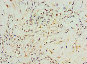
. AGR2 was detected in a paraffin-embedded section of human colorectal adenocarcinoma tissue. Heat mediated antigen retrieval was performed in EDTA buffer (pH 8.0, epitope retrieval solution). The tissue section was blocked with 10% goat serum. The tissue section was then incubated with 2 microg/ml rabbit anti-AGR2 Antibody (A02922-2) overnight at 4°C. Peroxidase Conjugated Goat Anti-rabbit IgG was used as secondary antibody and incubated for 30 minutes at 37°C. The tissue section was developed using HRP Conjugated Rabbit IgG Super Vision Assay Kit (Catalog # SV0002) with DAB as the chromogen.")
. AGR2 was detected in a paraffin-embedded section of human non-small cell lung cancer tissue. Heat mediated antigen retrieval was performed in EDTA buffer (pH 8.0, epitope retrieval solution). The tissue section was blocked with 10% goat serum. The tissue section was then incubated with 2 microg/ml rabbit anti-AGR2 Antibody (A02922-2) overnight at 4°C. Peroxidase Conjugated Goat Anti-rabbit IgG was used as secondary antibody and incubated for 30 minutes at 37°C. The tissue section was developed using HRP Conjugated Rabbit IgG Super Vision Assay Kit (Catalog # SV0002) with DAB as the chromogen.")
. AGR2 was detected in a paraffin-embedded section of human bladder infiltrating urothelial carcinoma with squamous differentiation tissue. Heat mediated antigen retrieval was performed in EDTA buffer (pH 8.0, epitope retrieval solution). The tissue section was blocked with 10% goat serum. The tissue section was then incubated with 2 microg/ml rabbit anti-AGR2 Antibody (A02922-2) overnight at 4°C. Peroxidase Conjugated Goat Anti-rabbit IgG was used as secondary antibody and incubated for 30 minutes at 37°C. The tissue section was developed using HRP Conjugated Rabbit IgG Super Vision Assay Kit (Catalog # SV0002) with DAB as the chromogen.")
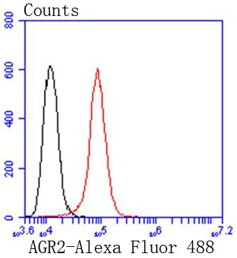
. Overlay histogram showing CACO-2 cells stained with A02922-2 (Blue line). The cells were fixed with 4% paraformaldehyde and blocked with 10% normal goat serum. And then incubated with rabbit anti-AGR2 Antibody (A02922-2, 1 microg/1x106 cells) for 30 min at 20°C. DyLight®488 conjugated goat anti-rabbit IgG (BA1127, 5-10 microg/1x106 cells) was used as secondary antibody for 30 minutes at 20°C. Isotype control antibody (Green line) was rabbit IgG (1 microg/1x106) used under the same conditions. Unlabelled sample (Red line) was also used as a control.")
Figure 1. Western blot analysis of AGR2 using anti-AGR2 antibody (A02922-2). Electrophoresis was performed on a 5-20% SDS-PAGE gel at 70V (Stacking gel) / 90V (Resolving gel) for 2-3 hours. The sample well of each lane was loaded with 30 ug of sample under reducing conditions. Lane 1: human T47D whole cell lysates, Lane 2: human A549 whole cell lysates, Lane 3: rat stomach tissue lysates, Lane 4: rat small intestine tissue lysates, Lane 5: mouse stomach tissue lysates, Lane 6: mouse small intestine tissue lysates. After electrophoresis, proteins were transferred to a nitrocellulose membrane at 150 mA for 50-90 minutes. Blocked the membrane with 5% non-fat milk/TBS for 1.5 hour at RT. The membrane was incubated with rabbit anti-AGR2 antigen affinity purified polyclonal antibody (Catalog # A02922-2) at 0.5 microg/mL overnight at 4°C, then washed with TBS-0.1%Tween 3 times with 5 minutes each and probed with a goat anti-rabbit IgG-HRP secondary antibody at a dilution of 1:5000 for 1.5 hour at RT. The signal is developed using an Enhanced Chemiluminescent detection (ECL) kit (Catalog # EK1002) with Tanon 5200 system. A specific band was detected for AGR2 at approximately 17 kDa. The expected band size for AGR2 is at 20 kDa.
Anti-Anterior Gradient 2/AGR2 Antibody Picoband(r)
A02922-2-CARRIER-FREE
ApplicationsFlow Cytometry, Western Blot, ImmunoHistoChemistry
Product group Antibodies
ReactivityHuman, Mouse, Rat
TargetAGR2
Overview
- SupplierBoster Bio
- Product NameAnti-Anterior Gradient 2/AGR2 Antibody Picoband(r)
- Delivery Days Customer9
- ApplicationsFlow Cytometry, Western Blot, ImmunoHistoChemistry
- CertificationResearch Use Only
- ClonalityPolyclonal
- Concentration500 ug/ml
- Gene ID10551
- Target nameAGR2
- Target descriptionanterior gradient 2, protein disulphide isomerase family member
- Target synonymsAG-2, AG2, GOB-4, HAG-2, HEL-S-116, HPC8, PDIA17, RIFTD, XAG-2, anterior gradient protein 2 homolog, anterior gradient homolog 2, epididymis secretory protein Li 116, protein disulfide isomerase family A, member 17, secreted cement gland homolog, secreted cement gland protein XAG-2 homolog
- HostRabbit
- IsotypeIgG
- Protein IDO95994
- Protein NameAnterior gradient protein 2 homolog
- Scientific DescriptionBoster Bio Anti-Anterior Gradient 2/AGR2 Antibody Picoband® catalog # A02922-2. Tested in Flow Cytometry, IHC, WB applications. This antibody reacts with Human, Mouse, Rat. The brand Picoband indicates this is a premium antibody that guarantees superior quality, high affinity, and strong signals with minimal background in Western blot applications. Only our best-performing antibodies are designated as Picoband, ensuring unmatched performance.
- ReactivityHuman, Mouse, Rat
- Storage Instruction-20°C,2°C to 8°C
- UNSPSC12352203
Related products
Product group Antibodies
AGR2 AntibodyCSB-PA001458ESR2HU
ApplicationsELISA, ImmunoHistoChemistry
ReactivityHuman
TargetAGR2
- SizePrice
Product group Antibodies

Anti-Arg-2 [CPR02]Ab03441-23.0
ApplicationsImmunoFluorescence, ELISA, Other Application
ReactivityHuman
TargetAGR2
- SizePrice
Product group Antibodies
Anti-AGR2 AntibodyA31848
ApplicationsWestern Blot, ImmunoHistoChemistry
ReactivityHuman, Mouse, Rat
- SizePrice
Product group Antibodies

Goat anti-AGR2EB07494
ApplicationsWestern Blot, ELISA
ReactivityCanine, Human, Mouse
TargetAGR2
- SizePrice

Product group Antibodies
Anti-AGR2 AntibodyHPA007912
ApplicationsWestern Blot, ImmunoCytoChemistry, ImmunoHistoChemistry
ReactivityHuman
TargetAGR2
- SizePrice
Product group Antibodies

AGR2 AntibodyLS-C400453
ApplicationsWestern Blot, ELISA, ImmunoHistoChemistry
ReactivityHuman, Mouse
TargetAGR2
- SizePrice
Product group Antibodies
ApplicationsImmunoPrecipitation, Western Blot, ImmunoCytoChemistry, ImmunoHistoChemistry
ReactivityMouse, Rat
TargetAGR2
- SizePrice
Product group Antibodies
AGR2 Recombinant AntibodyBSM-52594R
ApplicationsFlow Cytometry, ImmunoFluorescence, Western Blot, ImmunoCytoChemistry, ImmunoHistoChemistry, ImmunoHistoChemistry Frozen, ImmunoHistoChemistry Paraffin
ReactivityHuman, Mouse, Rat
TargetAGR2
- SizePrice