

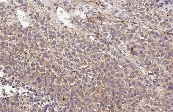
AGTR1 antibody [HL2524] detects AGTR1 protein at cell membrane and cytoplasm by immunohistochemical analysis. Sample: Paraffin-embedded Huh-7 xenograft. AGTR1 stained by AGTR1 antibody [HL2524] (GTX638885) diluted at 1:100. Antigen Retrieval: Citrate buffer, pH 6.0, 15 min
![Huh-7 cytoplasma extracts and membrane extracts (30 μg) were separated by 10% SDS-PAGE, and the membrane was blotted with AGTR1 antibody [HL2524] (GTX638885) diluted at 1:500. The HRP-conjugated anti-rabbit IgG antibody (GTX213110-01) was used to detect the primary antibody, and the signal was developed with Trident ECL plus-Enhanced. (CE: cytoplasma extract; ME: membrane extract)](https://www.genetex.com/upload/website/prouct_img/normal/GTX638885/GTX638885_T-45117_20230915_WB_Fraction_23091901_531.webp "Huh-7 cytoplasma extracts and membrane extracts (30 μg) were separated by 10% SDS-PAGE, and the membrane was blotted with AGTR1 antibody [HL2524] (GTX638885) diluted at 1:500. The HRP-conjugated anti-rabbit IgG antibody (GTX213110-01) was used to detect the primary antibody, and the signal was developed with Trident ECL plus-Enhanced. (CE: cytoplasma extract; ME: membrane extract)")
![Various whole cell extracts (30 μg) were separated by 10% SDS-PAGE, and the membrane was blotted with AGTR1 antibody [HL2524] (GTX638885) diluted at 1:1000. The HRP-conjugated anti-rabbit IgG antibody (GTX213110-01) was used to detect the primary antibody, and the signal was developed with Trident ECL plus-Enhanced. Corresponding RNA expression data for the same cell lines are based on Human Protein Atlas program.](https://www.genetex.com/upload/website/prouct_img/normal/GTX638885/GTX638885_45201_20231027_WB_TPM_watermark_23103019_803.webp "Various whole cell extracts (30 μg) were separated by 10% SDS-PAGE, and the membrane was blotted with AGTR1 antibody [HL2524] (GTX638885) diluted at 1:1000. The HRP-conjugated anti-rabbit IgG antibody (GTX213110-01) was used to detect the primary antibody, and the signal was developed with Trident ECL plus-Enhanced. Corresponding RNA expression data for the same cell lines are based on Human Protein Atlas program.")
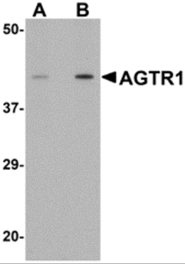
![Whole cell extract (30 μg) was separated by 10% SDS-PAGE, and the membrane was blotted with AGTR1 antibody [HL2524] (GTX638885) diluted at 1:1000. The HRP-conjugated anti-rabbit IgG antibody (GTX213110-01) was used to detect the primary antibody.](https://www.genetex.com/upload/website/prouct_img/normal/GTX638885/GTX638885_45201_20240105_WB_24010821_975.webp "Whole cell extract (30 μg) was separated by 10% SDS-PAGE, and the membrane was blotted with AGTR1 antibody [HL2524] (GTX638885) diluted at 1:1000. The HRP-conjugated anti-rabbit IgG antibody (GTX213110-01) was used to detect the primary antibody.")
![Whole cell extract (30 μg) was separated by 10% SDS-PAGE, and the membrane was blotted with AGTR1 antibody [HL2524] (GTX638885) diluted at 1:1000. The HRP-conjugated anti-rabbit IgG antibody (GTX213110-01) was used to detect the primary antibody, and the signal was developed with Trident femto Western HRP Substrate.](https://www.genetex.com/upload/website/prouct_img/normal/GTX638885/GTX638885_45201_20240105_WB_M_24010821_914.webp "Whole cell extract (30 μg) was separated by 10% SDS-PAGE, and the membrane was blotted with AGTR1 antibody [HL2524] (GTX638885) diluted at 1:1000. The HRP-conjugated anti-rabbit IgG antibody (GTX213110-01) was used to detect the primary antibody, and the signal was developed with Trident femto Western HRP Substrate.")
![AGTR1 antibody [HL2524] detects AGTR1 protein at cell membrane by immunofluorescent analysis. Sample: Huh-7 cells were fixed in 4% paraformaldehyde at RT for 15 min. Green: AGTR1 stained by AGTR1 antibody [HL2524] (GTX638885) diluted at 1:500. Red: alpha Tubulin, a cytoskeleton marker, stained by alpha Tubulin antibody [GT114] (GTX628802) diluted at 1:1000. Blue: Fluoroshield with DAPI (GTX30920).](https://www.genetex.com/upload/website/prouct_img/normal/GTX638885/GTX638885_45201_20240301_ICC_IF_24030600_536.webp "AGTR1 antibody [HL2524] detects AGTR1 protein at cell membrane by immunofluorescent analysis. Sample: Huh-7 cells were fixed in 4% paraformaldehyde at RT for 15 min. Green: AGTR1 stained by AGTR1 antibody [HL2524] (GTX638885) diluted at 1:500. Red: alpha Tubulin, a cytoskeleton marker, stained by alpha Tubulin antibody [GT114] (GTX628802) diluted at 1:1000. Blue: Fluoroshield with DAPI (GTX30920).")
![AGTR1 antibody [HL2524] detects AGTR1 protein by immunofluorescent analysis. Sample: Huh-7 cells were fixed in 4% paraformaldehyde at RT for 15 min. Green: AGTR1 stained by AGTR1 antibody [HL2524] (GTX638885) diluted at 1:500 and competitor's antibody (competitor) diluted at 1:500. Red: alpha Tubulin, a cytoskeleton marker, stained by alpha Tubulin antibody [GT114] (GTX628802) diluted at 1:1000. Blue: Fluoroshield with DAPI (GTX30920). *Competitor's antibody is not affiliated with GeneTex and does not endorse this product.](https://www.genetex.com/upload/website/prouct_img/normal/GTX638885/GTX638885_45201_20240419_ICC_IF_competitor_24051400_210.webp "AGTR1 antibody [HL2524] detects AGTR1 protein by immunofluorescent analysis. Sample: Huh-7 cells were fixed in 4% paraformaldehyde at RT for 15 min. Green: AGTR1 stained by AGTR1 antibody [HL2524] (GTX638885) diluted at 1:500 and competitor's antibody (competitor) diluted at 1:500. Red: alpha Tubulin, a cytoskeleton marker, stained by alpha Tubulin antibody [GT114] (GTX628802) diluted at 1:1000. Blue: Fluoroshield with DAPI (GTX30920). *Competitor's antibody is not affiliated with GeneTex and does not endorse this product.")
![AGTR1 antibody [HL2524] detects AGTR1 protein by immunohistochemical analysis. Sample: Paraffin-embedded mouse kidney. AGTR1 stained by AGTR1 antibody [HL2524] (GTX638885) diluted at 1:100. Antigen Retrieval: Citrate buffer, pH 6.0, 15 min](https://www.genetex.com/upload/website/prouct_img/normal/GTX638885/GTX638885_T-45117_20240620_IHC-P_M_24062501_573.webp "AGTR1 antibody [HL2524] detects AGTR1 protein by immunohistochemical analysis. Sample: Paraffin-embedded mouse kidney. AGTR1 stained by AGTR1 antibody [HL2524] (GTX638885) diluted at 1:100. Antigen Retrieval: Citrate buffer, pH 6.0, 15 min")
AGTR1 antibody [HL2524] detects AGTR1 protein at cell membrane and cytoplasm by immunohistochemical analysis. Sample: Paraffin-embedded Huh-7 xenograft. AGTR1 stained by AGTR1 antibody [HL2524] (GTX638885) diluted at 1:100. Antigen Retrieval: Citrate buffer, pH 6.0, 15 min
AGTR1 antibody [HL2524]
GTX638885
ApplicationsImmunoFluorescence, Western Blot, ImmunoCytoChemistry, ImmunoHistoChemistry, ImmunoHistoChemistry Paraffin
Product group Antibodies
ReactivityHuman, Mouse
TargetAGTR1
Overview
- SupplierGeneTex
- Product NameAGTR1 antibody [HL2524]
- Delivery Days Customer9
- Application Supplier NoteIHC-P: 1:100-1:1000. *Optimal dilutions/concentrations should be determined by the researcher.Not tested in other applications.
- ApplicationsImmunoFluorescence, Western Blot, ImmunoCytoChemistry, ImmunoHistoChemistry, ImmunoHistoChemistry Paraffin
- CertificationResearch Use Only
- ClonalityMonoclonal
- Clone IDHL2524
- Concentration1 mg/ml
- ConjugateUnconjugated
- Gene ID185
- Target nameAGTR1
- Target descriptionangiotensin II receptor type 1
- Target synonymsAG2S, AGTR1B, AT1, AT1AR, AT1B, AT1BR, AT1R, AT2R1, ATR1, HAT1R, type-1 angiotensin II receptor, AT1 receptor, type-1B angiotensin II receptor
- HostRabbit
- IsotypeIgG
- Protein IDP30556
- Protein NameType-1 angiotensin II receptor
- Scientific DescriptionAngiotensin II is a potent vasopressor hormone and a primary regulator of aldosterone secretion. It is an important effector controlling blood pressure and volume in the cardiovascular system. It acts through at least two types of receptors. This gene encodes the type 1 receptor which is thought to mediate the major cardiovascular effects of angiotensin II. This gene may play a role in the generation of reperfusion arrhythmias following restoration of blood flow to ischemic or infarcted myocardium. It was previously thought that a related gene, denoted as AGTR1B, existed; however, it is now believed that there is only one type 1 receptor gene in humans. Multiple alternatively spliced transcript variants have been reported for this gene. [provided by RefSeq, Jul 2012]
- ReactivityHuman, Mouse
- Storage Instruction-20°C or -80°C,2°C to 8°C
- UNSPSC41116161
Datasheet
Related products
Product group Antibodies
ApplicationsWestern Blot, ELISA
ReactivityHuman, Mouse
- SizePrice
Product group Antibodies
Anti-AGTR1 Antibody144-63128
ApplicationsWestern Blot
ReactivityHuman, Mouse, Rat
TargetAGTR1
- SizePrice
Product group Antibodies

AGTR1 / AT1 Receptor AntibodyLS-C749212
ApplicationsWestern Blot
ReactivityHuman, Mouse, Rat
TargetAGTR1
- SizePrice
Product group Antibodies
References
ApplicationsFlow Cytometry, ImmunoFluorescence, Western Blot, ELISA, ImmunoCytoChemistry, ImmunoHistoChemistry, ImmunoHistoChemistry Frozen, ImmunoHistoChemistry Paraffin
ReactivityHuman, Mouse, Rat
TargetAGTR1
- SizePrice
Product group Antibodies
AGTR1 AntibodyCSB-PA001465LA01HU
ApplicationsImmunoFluorescence, Western Blot, ELISA, ImmunoHistoChemistry
ReactivityHuman, Mouse
TargetAGTR1
- SizePrice
Product group Antibodies

Goat anti-AGTR1 / AT1EB07446
ApplicationsWestern Blot, ELISA
ReactivityHuman, Mouse
TargetAGTR1
- SizePrice
Product group Antibodies
Agtr1 Polyclonal AntibodyCAC07759
ApplicationsImmunoFluorescence, Western Blot, ELISA, ImmunoHistoChemistry
ReactivityMouse
TargetAGTR1
- SizePrice
Product group Antibodies
AGTR1 antibodyGTX31793
ApplicationsWestern Blot, ELISA, ImmunoHistoChemistry, ImmunoHistoChemistry Paraffin
ReactivityHuman, Mouse, Rat
TargetAGTR1
- SizePrice
Product group Antibodies
References
AGTR1 antibody, InternalGTX89149
ApplicationsWestern Blot
ReactivityHuman, Mouse
TargetAGTR1
- SizePrice