

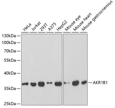
WB analysis of various sample lysates using GTX30040 AKR1B1 antibody. Dilution : 1:1000 Loading : 25μg per lane
WB analysis of various sample lysates using GTX30040 AKR1B1 antibody. Dilution : 1:1000 Loading : 25μg per lane
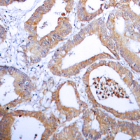
AKR1B1 antibody
GTX30040
ApplicationsWestern Blot
Product group Antibodies
ReactivityHuman, Mouse
TargetAKR1B1
Overview
- SupplierGeneTex
- Product NameAKR1B1 antibody
- Delivery Days Customer7
- Application Supplier NoteWB: 1:500 - 1:2000. *Optimal dilutions/concentrations should be determined by the researcher.Not tested in other applications.
- ApplicationsWestern Blot
- CertificationResearch Use Only
- ClonalityPolyclonal
- ConjugateUnconjugated
- Gene ID231
- Target nameAKR1B1
- Target descriptionaldo-keto reductase family 1 member B
- Target synonymsADR, ALDR1, ALR2, AR, aldo-keto reductase family 1 member B1, Lii5-2 CTCL tumor antigen, aldehyde reductase 1, aldose reductase, low Km aldose reductase
- HostRabbit
- IsotypeIgG
- Protein IDP15121
- Protein NameAldo-keto reductase family 1 member B1
- Scientific DescriptionThis gene encodes a member of the aldo/keto reductase superfamily, which consists of more than 40 known enzymes and proteins. This member catalyzes the reduction of a number of aldehydes, including the aldehyde form of glucose, and is thereby implicated in the development of diabetic complications by catalyzing the reduction of glucose to sorbitol. Multiple pseudogenes have been identified for this gene. The nomenclature system used by the HUGO Gene Nomenclature Committee to define human aldo-keto reductase family members is known to differ from that used by the Mouse Genome Informatics database. [provided by RefSeq, Feb 2009]
- ReactivityHuman, Mouse
- Storage Instruction-20°C or -80°C,2°C to 8°C
- UNSPSC41116161
References
- Protection of 6-OHDA neurotoxicity by PGF2alpha through FP-ERK-Nrf2 signaling in SH-SY5Y cells. Sano A et al., 2021 Feb 28, ToxicologyRead this paper
Datasheet
Related products
Product group Antibodies
Anti-AKR1B1 AntibodyA99317
ApplicationsWestern Blot, ELISA
ReactivityHuman, Rat
- SizePrice
Product group Antibodies
Anti-AKR1B1 Antibody144-60529
ApplicationsWestern Blot, ImmunoHistoChemistry
ReactivityHuman, Mouse, Rat
TargetAKR1B1
- SizePrice
Product group Antibodies
AKR1B1 Polyclonal AntibodyBS-2405R
ApplicationsImmunoFluorescence, Western Blot, ELISA, ImmunoCytoChemistry, ImmunoHistoChemistry, ImmunoHistoChemistry Frozen, ImmunoHistoChemistry Paraffin
ReactivityBovine, Canine, Human, Mouse, Rabbit, Rat
TargetAKR1B1
- SizePrice
Product group Antibodies
AKR1B1 AntibodyCSB-PA001539LA01HU
ApplicationsImmunoFluorescence, Western Blot, ELISA, ImmunoHistoChemistry
ReactivityHuman
TargetAKR1B1
- SizePrice
Product group Antibodies
AKR1B1 Polyclonal AntibodyCAC14579
ApplicationsImmunoFluorescence, Western Blot, ELISA, ImmunoHistoChemistry
TargetAKR1B1
- SizePrice

Product group Antibodies
Anti-AKR1B1 AntibodyHPA026425
ApplicationsImmunoCytoChemistry, ImmunoHistoChemistry
ReactivityHuman
TargetAKR1B1
- SizePrice
![Various whole cell extracts (30 μg) were separated by 10% SDS-PAGE, and the membrane was blotted with AKR1B1 antibody [N1C3] (GTX113381) diluted at 1:1000. The HRP-conjugated anti-rabbit IgG antibody (GTX213110-01) was used to detect the primary antibody. Corresponding RNA expression data for the same cell lines are based on Human Protein Atlas program.](https://www.genetex.com/upload/website/prouct_img/normal/GTX113381/GTX113381_43936_20200501_WB_TPM_watermark_w_23060501_162.webp)
Product group Antibodies
AKR1B1 antibody [N1C3]GTX113381
ApplicationsImmunoFluorescence, Western Blot, ImmunoCytoChemistry, ImmunoHistoChemistry, ImmunoHistoChemistry Paraffin
ReactivityHuman, Porcine, Rat
TargetAKR1B1
- SizePrice
Product group Antibodies
AKR1B1 antibodyGTX54911
ApplicationsImmunoFluorescence, Western Blot, ImmunoCytoChemistry, ImmunoHistoChemistry, ImmunoHistoChemistry Paraffin
ReactivityHuman, Mouse, Rat
TargetAKR1B1
- SizePrice
Product group Antibodies

AKR1B1 / Aldose Reductase AntibodyLS-C400454
ApplicationsWestern Blot, ELISA, ImmunoHistoChemistry
ReactivityHuman, Mouse, Rat
TargetAKR1B1
- SizePrice