


Anti-AKR1B1 Antibody
A99317
ApplicationsWestern Blot, ELISA
Product group Antibodies
ReactivityHuman, Rat
Overview
- SupplierAntibodies.com
- Product NameAnti-AKR1B1 Antibody
- Delivery Days Customer7
- ApplicationsWestern Blot, ELISA
- CertificationResearch Use Only
- ClonalityPolyclonal
- ConjugateUnconjugated
- HostRabbit
- IsotypeIgG
- Scientific DescriptionRabbit polyclonal antibody to AKR1B1.
- ReactivityHuman, Rat
- UNSPSC12352203
Related products
Product group Antibodies
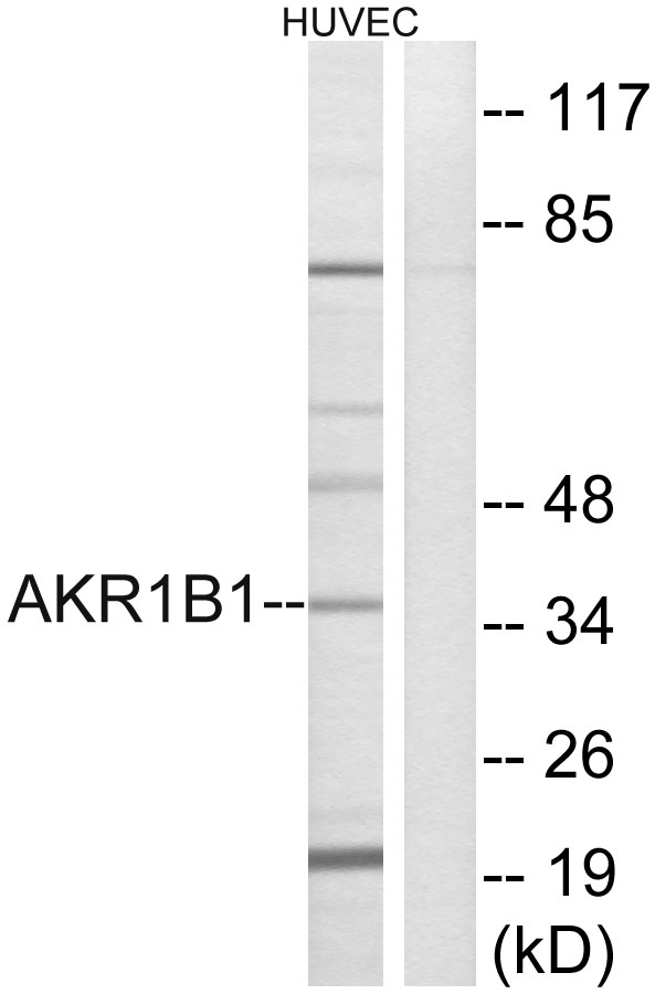
Anti-AKR1B1 Antibody144-60529
ApplicationsWestern Blot, ImmunoHistoChemistry
ReactivityHuman, Mouse, Rat
TargetAKR1B1
- SizePrice
Product group Antibodies
AKR1B1 Polyclonal AntibodyBS-2405R
ApplicationsImmunoFluorescence, Western Blot, ELISA, ImmunoCytoChemistry, ImmunoHistoChemistry, ImmunoHistoChemistry Frozen, ImmunoHistoChemistry Paraffin
ReactivityBovine, Canine, Human, Mouse, Rabbit, Rat
TargetAKR1B1
- SizePrice
Product group Antibodies
AKR1B1 AntibodyCSB-PA001539LA01HU
ApplicationsImmunoFluorescence, Western Blot, ELISA, ImmunoHistoChemistry
ReactivityHuman
TargetAKR1B1
- SizePrice
Product group Antibodies
AKR1B1 Polyclonal AntibodyCAC14579
ApplicationsImmunoFluorescence, Western Blot, ELISA, ImmunoHistoChemistry
TargetAKR1B1
- SizePrice

Product group Antibodies
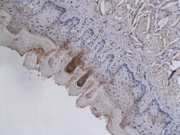
Anti-AKR1B1 AntibodyHPA026425
ApplicationsImmunoCytoChemistry, ImmunoHistoChemistry
ReactivityHuman
TargetAKR1B1
- SizePrice
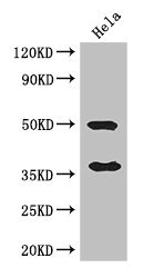
![Various whole cell extracts (30 μg) were separated by 10% SDS-PAGE, and the membrane was blotted with AKR1B1 antibody [N1C3] (GTX113381) diluted at 1:1000. The HRP-conjugated anti-rabbit IgG antibody (GTX213110-01) was used to detect the primary antibody. Corresponding RNA expression data for the same cell lines are based on Human Protein Atlas program.](https://www.genetex.com/upload/website/prouct_img/normal/GTX113381/GTX113381_43936_20200501_WB_TPM_watermark_w_23060501_162.webp)
Product group Antibodies
AKR1B1 antibody [N1C3]GTX113381
ApplicationsImmunoFluorescence, Western Blot, ImmunoCytoChemistry, ImmunoHistoChemistry, ImmunoHistoChemistry Paraffin
ReactivityHuman, Porcine, Rat
TargetAKR1B1
- SizePrice
Product group Antibodies

AKR1B1 / Aldose Reductase AntibodyLS-C400454
ApplicationsWestern Blot, ELISA, ImmunoHistoChemistry
ReactivityHuman, Mouse, Rat
TargetAKR1B1
- SizePrice

Product group Antibodies
Anti-AKR1B1 Antibody Picoband(r)PB10035-CARRIER-FREE
ApplicationsFlow Cytometry, Western Blot, ImmunoHistoChemistry
ReactivityHuman, Mouse, Rat
TargetAKR1B1
- SizePrice
Product group Antibodies
Anti-AKR1B1 AntibodyCAB13944
ApplicationsWestern Blot, ELISA
ReactivityHuman
TargetAKR1B1
- SizePrice