

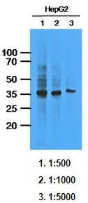
WB analysis of HepG2 lysate (33ug per lane) using AKR1C1 antibody at the indicated dilutions.
WB analysis of HepG2 lysate (33ug per lane) using AKR1C1 antibody at the indicated dilutions.
AKR1C1 antibody [AT6D10]
GTX53684
ApplicationsWestern Blot, ELISA
Product group Antibodies
ReactivityHuman
TargetAKR1C1
Overview
- SupplierGeneTex
- Product NameAKR1C1 antibody [AT6D10]
- Delivery Days Customer9
- Application Supplier NoteThe antibody has been tested by ELISA and Western blot analysis to assure specificity and reactivity. Since application varies, however, each investigation should be titrated by the reagent to obtain optimal results. Recommended dilution range for Western blot analysis is 1:1000. Recommended starting dilution is 1:1000
- ApplicationsWestern Blot, ELISA
- CertificationResearch Use Only
- ClonalityMonoclonal
- Clone IDAT6D10
- Concentration1 mg/ml
- ConjugateUnconjugated
- Gene ID1645
- Target nameAKR1C1
- Target descriptionaldo-keto reductase family 1 member C1
- Target synonyms2-ALPHA-HSD, 20-ALPHA-HSD, C9, DD1, DD1/DD2, DDH, DDH1, H-37, HAKRC, HBAB, MBAB, aldo-keto reductase family 1 member C1, 20 alpha-hydroxysteroid dehydrogenase, aldo-keto reductase C, chlordecone reductase homolog HAKRC, dihydrodiol dehydrogenase 1, dihydrodiol dehydrogenase 1/2, dihydrodiol dehydrogenase 1; 20-alpha (3-alpha)-hydroxysteroid dehydrogenase, hepatic dihydrodiol dehydrogenase, high-affinity hepatic bile acid-binding protein, indanol dehydrogenase, trans-1,2-dihydrobenzene-1,2-diol dehydrogenase, type II 3-alpha-hydroxysteroid dehydrogenase
- HostMouse
- IsotypeIgG1
- Protein IDQ04828
- Protein NameAldo-keto reductase family 1 member C1
- Scientific DescriptionThis gene encodes a member of the aldo/keto reductase superfamily, which consists of more than 40 known enzymes and proteins. These enzymes catalyze the conversion of aldehydes and ketones to their corresponding alcohols by utilizing NADH and/or NADPH as cofactors. The enzymes display overlapping but distinct substrate specificity. This enzyme catalyzes the reaction of progesterone to the inactive form 20-alpha-hydroxy-progesterone. This gene shares high sequence identity with three other gene members and is clustered with those three genes at chromosome 10p15-p14. [provided by RefSeq, Jul 2008]
- ReactivityHuman
- Storage Instruction-20°C or -80°C,2°C to 8°C
- UNSPSC41116161
References
- Transcriptional consequences of XPA disruption in human cell lines. Manandhar M et al., 2017 Sep, DNA Repair (Amst)Read this paper
Datasheet
Related products
Product group Antibodies
Anti-AKR1C1 AntibodyA28765
ApplicationsWestern Blot
ReactivityHuman, Mouse, Rat
- SizePrice
Product group Antibodies
Anti-AKR1C1 Antibody144-60143
ApplicationsWestern Blot
ReactivityHuman, Mouse
TargetAKR1C1
- SizePrice
Product group Antibodies
AKR1C1/2 Recombinant AntibodyBSM-62246R
ApplicationsFlow Cytometry, Western Blot
ReactivityHuman, Mouse, Rat
TargetAKR1C1
- SizePrice
Product group Antibodies
AKR1C1 AntibodyCSB-PA001542LA01HU
ApplicationsImmunoFluorescence, ELISA
ReactivityHuman
TargetAKR1C1
- SizePrice
Product group Antibodies

DDH / AKR1C1 AntibodyLS-C402612
ApplicationsWestern Blot, ELISA, ImmunoHistoChemistry
ReactivityHuman
TargetAKR1C1
- SizePrice

Product group Antibodies
Anti-AKR1C1 AntibodyHPA068265
ApplicationsWestern Blot, ImmunoCytoChemistry
ReactivityHuman
TargetAKR1C1
- SizePrice
Product group Antibodies
AKR1C1 antibodyGTX105620
ApplicationsImmunoFluorescence, Western Blot, ImmunoCytoChemistry, ImmunoHistoChemistry, ImmunoHistoChemistry Paraffin
ReactivityHuman, Mouse
TargetAKR1C1
- SizePrice
Product group Antibodies
Anti-AKR1C1Y058805
ApplicationsWestern Blot, ImmunoHistoChemistry
ReactivityHuman
- SizePrice