

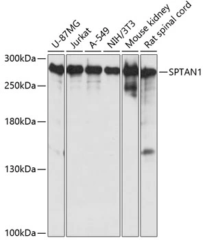
WB analysis of various sample lysates using GTX66033 alpha Fodrin antibody. Dilution : 1:3000 Loading : 25μg per lane


WB analysis of various sample lysates using GTX66033 alpha Fodrin antibody. Dilution : 1:3000 Loading : 25μg per lane
alpha Fodrin antibody
GTX66033
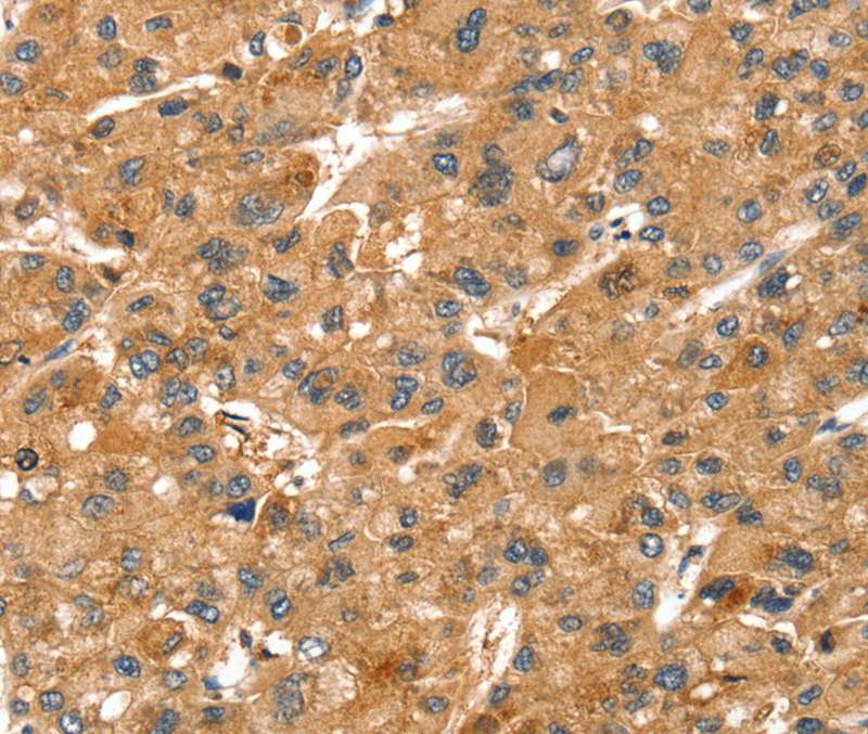
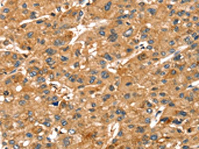
ApplicationsImmunoFluorescence, Western Blot, ImmunoCytoChemistry, ImmunoHistoChemistry, ImmunoHistoChemistry Paraffin
Product group Antibodies
ReactivityHuman, Mouse, Rat
TargetSPTAN1
Overview
- SupplierGeneTex
- Product Namealpha Fodrin antibody
- Delivery Days Customer9
- Application Supplier NoteWB: 1:500 - 1:2000. ICC/IF: 1:50 - 1:200. IHC-P: 1:50 - 1:200. *Optimal dilutions/concentrations should be determined by the researcher.Not tested in other applications.
- ApplicationsImmunoFluorescence, Western Blot, ImmunoCytoChemistry, ImmunoHistoChemistry, ImmunoHistoChemistry Paraffin
- CertificationResearch Use Only
- ClonalityPolyclonal
- ConjugateUnconjugated
- Gene ID6709
- Target nameSPTAN1
- Target descriptionspectrin alpha, non-erythrocytic 1
- Target synonymsDEE5, DEVEP, EIEE5, HMN11, HMND11, NEAS, SPG91, SPTA2, spectrin alpha chain, non-erythrocytic 1, alpha-II spectrin, alpha-fodrin, epididymis secretory sperm binding protein, fodrin alpha chain, spectrin, non-erythroid alpha chain, spectrin, non-erythroid alpha subunit
- HostRabbit
- IsotypeIgG
- Protein IDQ13813
- Protein NameSpectrin alpha chain, non-erythrocytic 1
- Scientific DescriptionSpectrins are a family of filamentous cytoskeletal proteins that function as essential scaffold proteins that stabilize the plasma membrane and organize intracellular organelles. Spectrins are composed of alpha and beta dimers that associate to form tetramers linked in a head-to-head arrangement. This gene encodes an alpha spectrin that is specifically expressed in nonerythrocytic cells. The encoded protein has been implicated in other cellular functions including DNA repair and cell cycle regulation. Mutations in this gene are the cause of early infantile epileptic encephalopathy-5. Alternate splicing results in multiple transcript variants.[provided by RefSeq, Sep 2010]
- ReactivityHuman, Mouse, Rat
- Storage Instruction-20°C or -80°C,2°C to 8°C
- UNSPSC41116161
Datasheet
Related products
Product group Antibodies
Anti-SPTAN1 AntibodyA45625
ApplicationsImmunoHistoChemistry
ReactivityHuman, Mouse, Rat
- SizePrice

Product group Antibodies
Anti-NEAS/SPTAN1 Antibody Picoband(r)A03831-3-CARRIER-FREE
ApplicationsFlow Cytometry, ImmunoFluorescence, Western Blot, ELISA, ImmunoCytoChemistry, ImmunoHistoChemistry
ReactivityHuman, Mouse, Rat
TargetSPTAN1
- SizePrice
Product group Antibodies
Anti-SPTAN1 Antibody144-60011
ApplicationsWestern Blot
ReactivityHuman, Mouse, Rat
TargetSPTAN1
- SizePrice
Product group Antibodies
SPECA Recombinant Antibody, AbBy Fluor-488 ConjugatedBSM-61537R-BF488
ApplicationsImmunoFluorescence, Western Blot
ReactivityHuman, Mouse, Rat
TargetSPTAN1
- SizePrice
Product group Antibodies
SPTAN1 AntibodyCSB-PA156898
ApplicationsELISA, ImmunoHistoChemistry
ReactivityHuman, Mouse, Rat
TargetSPTAN1
- SizePrice
Product group Antibodies
ApplicationsImmunoPrecipitation, Western Blot, ImmunoCytoChemistry, ImmunoHistoChemistry
ReactivityMouse, Rat
TargetSPTAN1
- SizePrice
Product group Antibodies

SPTAN1 / Alpha Fodrin AntibodyLS-C403608
ApplicationsELISA, ImmunoHistoChemistry
ReactivityHuman, Mouse, Rat
TargetSPTAN1
- SizePrice
Product group Antibodies
ApplicationsWestern Blot
ReactivityHuman
TargetSPTAN1
- SizePrice

Product group Antibodies
Anti-SPTAN1 AntibodyHPA007927
ApplicationsWestern Blot, ImmunoCytoChemistry, ImmunoHistoChemistry
ReactivityHuman
TargetSPTAN1
- SizePrice