

AMACR / p504S (Prostate Cancer Marker)(13H4), Biotin conjugate, 0.1mg/mL [26628-22-8]
BNCB0664
ReactivityBovine, Human, Mouse
Product group Antibodies
TargetAMACR
Overview
- SupplierBiotium
- Product NameAMACR / p504S (Prostate Cancer Marker)(13H4), Biotin conjugate, 0.1mg/mL [26628-22-8]
- Delivery Days Customer9
- CAS Number26628-22-8
- CertificationResearch Use Only
- ClonalityMonoclonal
- Clone ID13h4
- Concentration0.1 mg/ml
- ConjugateBiotin
- Gene ID23600
- Target nameAMACR
- Target descriptionalpha-methylacyl-CoA racemase
- Target synonymsAMACRD, CBAS4, P504S, RACE, RM, alpha-methylacyl-CoA racemase, 2-methylacyl-CoA racemase
- HostMouse
- IsotypeIgG
- Protein IDQ9UHK6
- Protein NameAlpha-methylacyl-CoA racemase
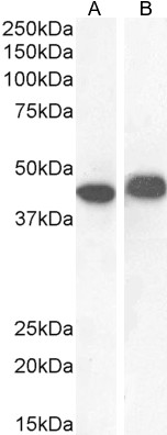
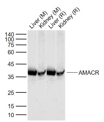
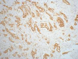
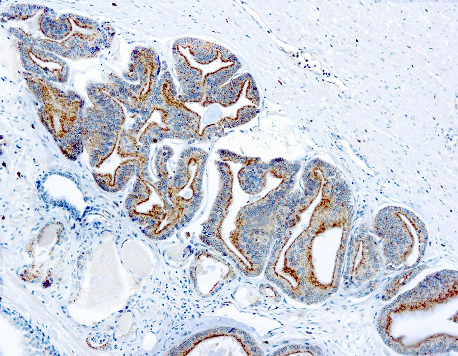
- Scientific DescriptionThis antibody recognizes a protein of 42 kDa, which is identified as Alpha-methylacyl-CoA Racemase (AMACR), also known as p504S. It is an enzyme that is involved in bile acid biosynthesis and -oxidation of branched-chain fatty acids. AMACR is essential in lipid metabolism. It is expressed in cells of premalignant high-grade prostatic intraepithelial neoplasia (HGPIN) and prostate adenocarcinoma. The majority of the carcinoma cells show a distinct granular cytoplasmic staining reaction. AMACR is present at low or undetectable levels in glandular epithelial cells of normal prostate and benign prostatic hyperplasia. A spotty granular cytoplasmic staining is seen in a few cells of the benign glands. AMACR is expressed in normal liver (hepatocytes), kidney (tubular epithelial cells) and gall bladder (epithelial cells). Expression has also been found in lung (bronchial epithelial cells) and colon (colonic surface epithelium). AMACR expression can also be found in hepatocellular carcinoma and kidney carcinoma. Past studies have also shown that AMACR is expressed in various colon carcinomas (well, moderately and poorly differentiated) and over expressed in prostate carcinoma.
- SourceAnimal
- ReactivityBovine, Human, Mouse
- Storage Instruction2°C to 8°C,RT
- UNSPSC41116161
MSDS
Related products
Product group Antibodies
Anti-AMACR AntibodyA82680
ApplicationsWestern Blot, ELISA, ImmunoHistoChemistry
ReactivityHuman
- SizePrice
Product group Antibodies
Anti-AMACR Antibody144-01130
ApplicationsWestern Blot
ReactivityHuman, Mouse
TargetAMACR
- SizePrice

Product group Antibodies
Anti-AMACR AntibodyAMAB91561
ApplicationsWestern Blot, ImmunoHistoChemistry
ReactivityHuman
TargetAMACR
- SizePrice
Product group Antibodies

AMACR / P504S Antibody (clone 1F1)LS-C764021
ApplicationsImmunoFluorescence, Western Blot, ImmunoHistoChemistry, ImmunoHistoChemistry Paraffin
ReactivityHuman, Mouse, Rat
TargetAMACR
- SizePrice
Product group Antibodies
AMACR Polyclonal AntibodyBS-0840R
ApplicationsWestern Blot, ELISA, ImmunoHistoChemistry, ImmunoHistoChemistry Frozen, ImmunoHistoChemistry Paraffin
ReactivityHuman, Mouse, Rat
TargetAMACR
- SizePrice
Product group Antibodies
AMACR Monoclonal AntibodyCSB-MA000203
ApplicationsWestern Blot, ELISA, ImmunoHistoChemistry
ReactivityHuman, Mouse, Rat
TargetAMACR
- SizePrice
Product group Antibodies

Goat anti-AMACREB12538
ApplicationsWestern Blot, ELISA
ReactivityHuman
TargetAMACR
- SizePrice
Product group Antibodies
Amacr Polyclonal AntibodyCAC08104
ApplicationsImmunoFluorescence, Western Blot, ELISA
ReactivityMouse
TargetAMACR
- SizePrice
Product group Antibodies
AMACR antibodyGTX12498
ApplicationsImmunoHistoChemistry, ImmunoHistoChemistry Paraffin
ReactivityHuman
TargetAMACR
- SizePrice