


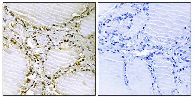
IHC-P analysis of human thyroid gland tissue using GTX86973 AMPD1 antibody. The picture on the right is blocked with the synthesized peptide.
IHC-P analysis of human thyroid gland tissue using GTX86973 AMPD1 antibody. The picture on the right is blocked with the synthesized peptide.
AMPD1 antibody
GTX86973
ApplicationsImmunoHistoChemistry, ImmunoHistoChemistry Paraffin
Product group Antibodies
ReactivityHuman
TargetAMPD1
Overview
- SupplierGeneTex
- Product NameAMPD1 antibody
- Delivery Days Customer9
- Application Supplier NoteIHC-P: 1:50~1:100. *Optimal dilutions/concentrations should be determined by the researcher.Not tested in other applications.
- ApplicationsImmunoHistoChemistry, ImmunoHistoChemistry Paraffin
- CertificationResearch Use Only
- ClonalityPolyclonal
- ConjugateUnconjugated
- Gene ID270
- Target nameAMPD1
- Target descriptionadenosine monophosphate deaminase 1
- Target synonymsMAD, MADA, MMDD, AMP deaminase 1, AMPD, adenosine monophosphate deaminase 1 (isoform M), adenosine monophosphate deaminase-1 (muscle), myoadenylate deaminase, skeletal muscle AMPD
- HostRabbit
- IsotypeIgG
- Protein IDP23109
- Protein NameAMP deaminase 1
- Scientific DescriptionAdenosine monophosphate deaminase 1 catalyzes the deamination of AMP to IMP in skeletal muscle and plays an important role in the purine nucleotide cycle. Two other genes have been identified, AMPD2 and AMPD3, for the liver- and erythocyte-specific isoforms, respectively. Deficiency of the muscle-specific enzyme is apparently a common cause of exercise-induced myopathy and probably the most common cause of metabolic myopathy in the human. Alternatively spliced transcript variants encoding different isoforms have been identified in this gene.[provided by RefSeq, Feb 2010]
- ReactivityHuman
- Storage Instruction-20°C or -80°C,2°C to 8°C
- UNSPSC41116161
Datasheet
Related products
Product group Antibodies
Anti-AMPD1 AntibodyA97051
ApplicationsELISA, ImmunoHistoChemistry
ReactivityHuman, Mouse, Rat
- SizePrice

Product group Antibodies
Anti-AMPD1 Antibody Picoband(r)A04209-1-CARRIER-FREE
ApplicationsFlow Cytometry, Western Blot, ELISA, ImmunoHistoChemistry
ReactivityHuman, Mouse, Rat
TargetAMPD1
- SizePrice
Product group Antibodies
Anti-AMPD1 Antibody144-03584
ApplicationsWestern Blot
ReactivityHuman, Mouse, Rat
TargetAMPD1
- SizePrice
Product group Antibodies
AMPD1 AntibodyCSB-PA001680LA01HU
ApplicationsWestern Blot, ELISA
ReactivityHuman
TargetAMPD1
- SizePrice
Product group Antibodies

Goat anti-AMPD1EB08258
ApplicationsELISA
ReactivityBovine, Canine, Human, Mouse, Porcine, Rat
TargetAMPD1
- SizePrice
Product group Antibodies
AMPD1 Polyclonal AntibodyCAC15036
ApplicationsWestern Blot, ELISA
TargetAMPD1
- SizePrice
Product group Antibodies

AMPD1 AntibodyLS-C402884
ApplicationsWestern Blot, ELISA, ImmunoHistoChemistry
ReactivityHuman
TargetAMPD1
- SizePrice

Product group Antibodies
Anti-AMPD1 AntibodyHPA028080
ApplicationsWestern Blot, ImmunoHistoChemistry
ReactivityHuman
TargetAMPD1
- SizePrice
Product group Antibodies
AMPD1 antibodyGTX55510
ApplicationsWestern Blot, ImmunoHistoChemistry, ImmunoHistoChemistry Paraffin
ReactivityHuman, Mouse, Rat
TargetAMPD1
- SizePrice
Product group Antibodies
Anti-AMPD1Y058345
ApplicationsELISA, ImmunoHistoChemistry
ReactivityHuman, Mouse, Rat
- SizePrice