

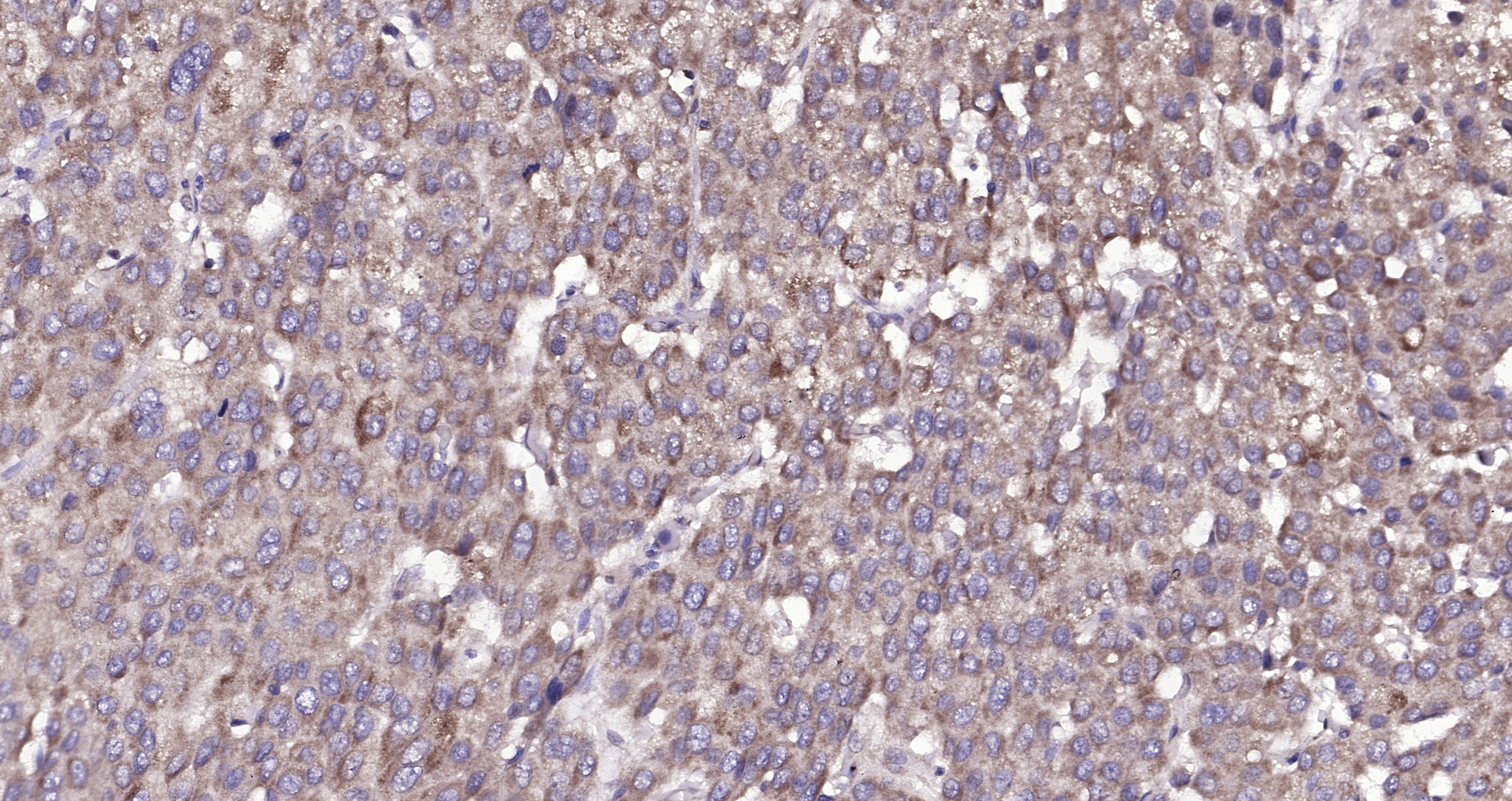
IHC-P analysis of human liver carcinoma tissue using GTX04890 Amphiregulin antibody. Antigen retrieval : Heat mediated sodium citrate, pH6.0 Dilution : 1:200


IHC-P analysis of human liver carcinoma tissue using GTX04890 Amphiregulin antibody. Antigen retrieval : Heat mediated sodium citrate, pH6.0 Dilution : 1:200
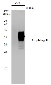
Amphiregulin antibody
GTX04890
ApplicationsImmunoFluorescence, Western Blot, ImmunoCytoChemistry, ImmunoHistoChemistry, ImmunoHistoChemistry Frozen, ImmunoHistoChemistry Paraffin
Product group Antibodies
ReactivityGuinea Pig, Human, Mouse, Rat
TargetAREG
Overview
- SupplierGeneTex
- Product NameAmphiregulin antibody
- Delivery Days Customer7
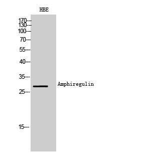
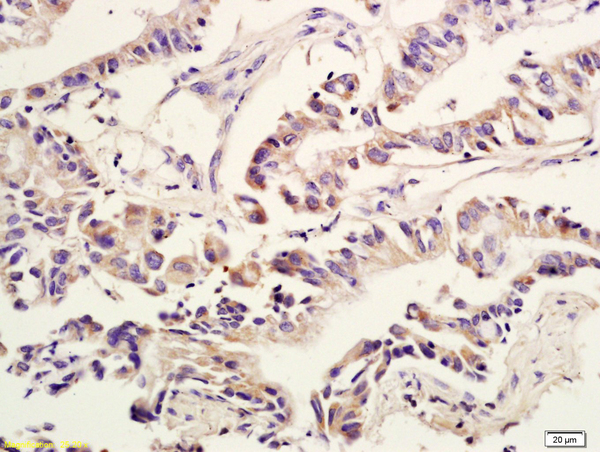
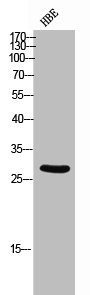
- Application Supplier NoteWB: 1:300-1:5000. ICC/IF: 1:50-200. IHC-P: 1:200-1:400. IHC-Fr: 1:50-200. *Optimal dilutions/concentrations should be determined by the researcher.Not tested in other applications.
- ApplicationsImmunoFluorescence, Western Blot, ImmunoCytoChemistry, ImmunoHistoChemistry, ImmunoHistoChemistry Frozen, ImmunoHistoChemistry Paraffin
- CertificationResearch Use Only
- ClonalityPolyclonal
- Concentration1 mg/ml
- ConjugateUnconjugated
- Gene ID374
- Target nameAREG
- Target descriptionamphiregulin
- Target synonymsAR, AREGB, CRDGF, SDGF, amphiregulin, amphiregulin B, colorectum cell-derived growth factor, schwannoma-derived growth factor
- HostRabbit
- IsotypeIgG
- Protein IDP15514
- Protein NameAmphiregulin
- Scientific DescriptionThe protein encoded by this gene is a member of the epidermal growth factor family. It is an autocrine growth factor as well as a mitogen for astrocytes, Schwann cells and fibroblasts. It is related to epidermal growth factor (EGF) and transforming growth factor alpha (TGF-alpha). The protein interacts with the EGF/TGF-alpha receptor to promote the growth of normal epithelial cells, and it inhibits the growth of certain aggressive carcinoma cell lines. It also functions in mammary gland, oocyte and bone tissue development. This gene is associated with a psoriasis-like skin phenotype, and is also associated with other pathological disorders, including various types of cancers and inflammatory conditions. [provided by RefSeq, Apr 2014]
- ReactivityGuinea Pig, Human, Mouse, Rat
- Storage Instruction-20°C or -80°C,2°C to 8°C
- UNSPSC41116161
Datasheet
Related products
Product group Antibodies
ApplicationsWestern Blot, ELISA
ReactivityHuman
- SizePrice
Product group Antibodies

AREG / Amphiregulin AntibodyLS-C747768
ApplicationsWestern Blot
ReactivityHuman, Mouse, Rat
TargetAREG
- SizePrice

Product group Antibodies
Anti-Amphiregulin/AREG Antibody Picoband(r)A01787-2-CARRIER-FREE
ApplicationsFlow Cytometry, Western Blot, ELISA
ReactivityHuman
TargetAREG
- SizePrice
Product group Antibodies
References
Amphiregulin Polyclonal AntibodyBS-3847R
ApplicationsFlow Cytometry, ImmunoFluorescence, Western Blot, ELISA, ImmunoCytoChemistry, ImmunoHistoChemistry, ImmunoHistoChemistry Frozen, ImmunoHistoChemistry Paraffin
ReactivityBovine, Canine, Equine, Human, Mouse, Rabbit, Rat, Sheep
TargetAREG
- SizePrice
Product group Antibodies
AREG AntibodyCSB-PA006314
ApplicationsWestern Blot, ELISA
ReactivityHuman
TargetAREG
- SizePrice
Product group Antibodies
Areg Polyclonal AntibodyCAC09140
ApplicationsImmunoFluorescence, ELISA, ImmunoHistoChemistry
TargetAREG
- SizePrice
Product group Antibodies
Amphiregulin antibodyGTX100986
ApplicationsWestern Blot, ImmunoHistoChemistry, ImmunoHistoChemistry Paraffin
ReactivityHuman
TargetAREG
- SizePrice

Product group Antibodies
Anti-AREG AntibodyHPA008720
ApplicationsImmunoHistoChemistry
ReactivityHuman
TargetAREG
- SizePrice
Product group Antibodies
Amphiregulin antibody [8K16]GTX52540
ApplicationsWestern Blot
ReactivityHuman
TargetAREG
- SizePrice

Product group Antibodies
Amphiregulin antibodyGTX55511
ApplicationsWestern Blot
ReactivityHuman, Mouse, Rat
TargetAREG
- SizePrice