

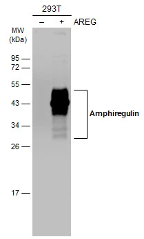
Non-transfected (–) and transfected (+) 293T whole cell extracts (30 μg) were separated by 12% SDS-PAGE, and the membrane was blotted with Amphiregulin antibody (GTX100986) diluted at 1:3000. The HRP-conjugated anti-rabbit IgG antibody (GTX213110-01) was used to detect the primary antibody.
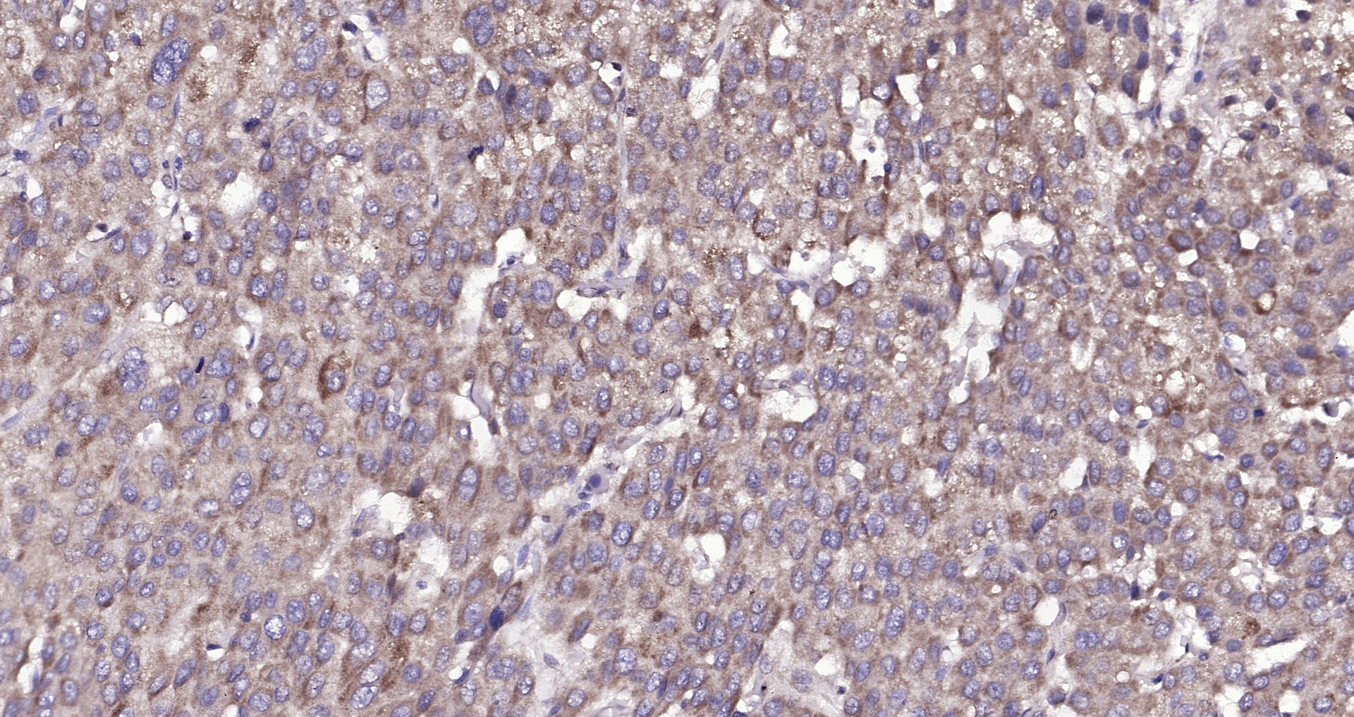
 diluted at 1:500.
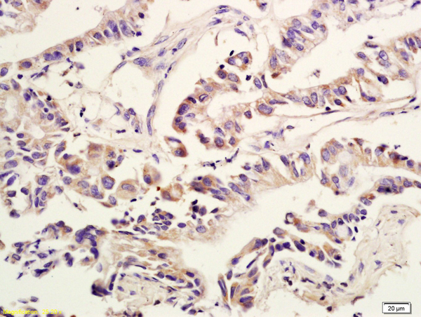
Antigen Retrieval: Citrate buffer, pH 6.0, 15 min")

Non-transfected (–) and transfected (+) 293T whole cell extracts (30 μg) were separated by 12% SDS-PAGE, and the membrane was blotted with Amphiregulin antibody (GTX100986) diluted at 1:3000. The HRP-conjugated anti-rabbit IgG antibody (GTX213110-01) was used to detect the primary antibody.
Amphiregulin antibody
GTX100986
ApplicationsWestern Blot, ImmunoHistoChemistry, ImmunoHistoChemistry Paraffin
Product group Antibodies
ReactivityHuman
TargetAREG
Overview
- SupplierGeneTex
- Product NameAmphiregulin antibody
- Delivery Days Customer9
- Application Supplier NoteWB: 1:500-1:3000. IHC-P: 1:100-1:1000. *Optimal dilutions/concentrations should be determined by the researcher.Not tested in other applications.
- ApplicationsWestern Blot, ImmunoHistoChemistry, ImmunoHistoChemistry Paraffin
- CertificationResearch Use Only
- ClonalityPolyclonal
- Concentration0.17 mg/ml
- ConjugateUnconjugated
- Gene ID374
- Target nameAREG
- Target descriptionamphiregulin
- Target synonymsAR, AREGB, CRDGF, SDGF, amphiregulin, amphiregulin B, colorectum cell-derived growth factor, schwannoma-derived growth factor
- HostRabbit
- IsotypeIgG
- Protein IDP15514
- Protein NameAmphiregulin
- Scientific DescriptionThe protein encoded by this gene is a member of the epidermal growth factor family. It is an autocrine growth factor as well as a mitogen for astrocytes, Schwann cells, and fibroblasts. It is related to epidermal growth factor (EGF) and transforming growth factor alpha (TGF-alpha). This protein interacts with the EGF/TGF-alpha receptor to promote the growth of normal epithelial cells and inhibits the growth of certain aggressive carcinoma cell lines. This encoded protein is associated with a psoriasis-like skin phenotype. [provided by RefSeq]
- ReactivityHuman
- Storage Instruction-20°C or -80°C,2°C to 8°C
- UNSPSC41116161
Datasheet
Related products
Product group Antibodies
ApplicationsWestern Blot, ELISA
ReactivityHuman
- SizePrice
Product group Antibodies

AREG / Amphiregulin AntibodyLS-C747768
ApplicationsWestern Blot
ReactivityHuman, Mouse, Rat
TargetAREG
- SizePrice

Product group Antibodies
Anti-Amphiregulin/AREG Antibody Picoband(r)A01787-2-CARRIER-FREE
ApplicationsFlow Cytometry, Western Blot, ELISA
ReactivityHuman
TargetAREG
- SizePrice
Product group Antibodies
References
Amphiregulin Polyclonal AntibodyBS-3847R
ApplicationsFlow Cytometry, ImmunoFluorescence, Western Blot, ELISA, ImmunoCytoChemistry, ImmunoHistoChemistry, ImmunoHistoChemistry Frozen, ImmunoHistoChemistry Paraffin
ReactivityBovine, Canine, Equine, Human, Mouse, Rabbit, Rat, Sheep
TargetAREG
- SizePrice
Product group Antibodies
AREG AntibodyCSB-PA006314
ApplicationsWestern Blot, ELISA
ReactivityHuman
TargetAREG
- SizePrice
Product group Antibodies
Areg Polyclonal AntibodyCAC09140
ApplicationsImmunoFluorescence, ELISA, ImmunoHistoChemistry
TargetAREG
- SizePrice
Product group Antibodies
Amphiregulin antibodyGTX04890
ApplicationsImmunoFluorescence, Western Blot, ImmunoCytoChemistry, ImmunoHistoChemistry, ImmunoHistoChemistry Frozen, ImmunoHistoChemistry Paraffin
ReactivityGuinea Pig, Human, Mouse, Rat
TargetAREG
- SizePrice

Product group Antibodies
Anti-AREG AntibodyHPA008720
ApplicationsImmunoHistoChemistry
ReactivityHuman
TargetAREG
- SizePrice
Product group Antibodies
Amphiregulin antibody [8K16]GTX52540
ApplicationsWestern Blot
ReactivityHuman
TargetAREG
- SizePrice

Product group Antibodies
Amphiregulin antibodyGTX55511
ApplicationsWestern Blot
ReactivityHuman, Mouse, Rat
TargetAREG
- SizePrice