


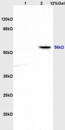
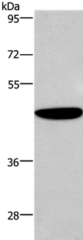
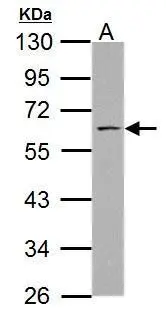
Figure 1. Western blot analysis of SSH3BP1/ABI1 using anti-SSH3BP1/ABI1 antibody (PB9416). Electrophoresis was performed on a 5-20% SDS-PAGE gel at 70V (Stacking gel) / 90V (Resolving gel) for 2-3 hours. The sample well of each lane was loaded with 30 ug of sample under reducing conditions. Lane 1: human 293T whole cell lysates, Lane 2: human MCF-7 whole cell lysates, Lane 3: human U87 whole cell lysates, Lane 4: rat brain tissue lysates, Lane 5: rat C6 whole cell lysates, Lane 6: mouse brain tissue lysates. After electrophoresis, proteins were transferred to a nitrocellulose membrane at 150 mA for 50-90 minutes. Blocked the membrane with 5% non-fat milk/TBS for 1.5 hour at RT. The membrane was incubated with rabbit anti-SSH3BP1/ABI1 antigen affinity purified polyclonal antibody (Catalog # PB9416) at 0.5 microg/mL overnight at 4°C, then washed with TBS-0.1%Tween 3 times with 5 minutes each and probed with a goat anti-rabbit IgG-HRP secondary antibody at a dilution of 1:5000 for 1.5 hour at RT. The signal is developed using an Enhanced Chemiluminescent detection (ECL) kit (Catalog # EK1002) with Tanon 5200 system. A specific band was detected for SSH3BP1/ABI1 at approximately 65 kDa. The expected band size for SSH3BP1/ABI1 is at 55 kDa.
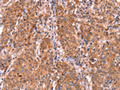
. SSH3BP1/ABI1 was detected in a paraffin-embedded section of mouse brain tissue. Heat mediated antigen retrieval was performed in EDTA buffer (pH 8.0, epitope retrieval solution). The tissue section was blocked with 10% goat serum. The tissue section was then incubated with 2 microg/ml rabbit anti-SSH3BP1/ABI1 Antibody (PB9416) overnight at 4°C. Peroxidase Conjugated Goat Anti-rabbit IgG was used as secondary antibody and incubated for 30 minutes at 37°C. The tissue section was developed using HRP Conjugated Rabbit IgG Super Vision Assay Kit (Catalog # SV0002) with DAB as the chromogen.")
. SSH3BP1/ABI1 was detected in a paraffin-embedded section of rat brain tissue. Heat mediated antigen retrieval was performed in EDTA buffer (pH 8.0, epitope retrieval solution). The tissue section was blocked with 10% goat serum. The tissue section was then incubated with 2 microg/ml rabbit anti-SSH3BP1/ABI1 Antibody (PB9416) overnight at 4°C. Peroxidase Conjugated Goat Anti-rabbit IgG was used as secondary antibody and incubated for 30 minutes at 37°C. The tissue section was developed using HRP Conjugated Rabbit IgG Super Vision Assay Kit (Catalog # SV0002) with DAB as the chromogen.")
. SSH3BP1/ABI1 was detected in an immunocytochemical section of A431 cells. Enzyme antigen retrieval was performed using IHC enzyme antigen retrieval reagent (AR0022) for 15 mins. The cells were blocked with 10% goat serum. And then incubated with 5 microg/mL rabbit anti-SSH3BP1/ABI1 Antibody (PB9416) overnight at 4°C. DyLight®488 Conjugated Goat Anti-Rabbit IgG (BA1127) was used as secondary antibody at 1:100 dilution and incubated for 30 minutes at 37°C. The section was counterstained with DAPI. Visualize using a fluorescence microscope and filter sets appropriate for the label used.")
. Overlay histogram showing K562 cells stained with PB9416 (Blue line). To facilitate intracellular staining, cells were fixed with 4% paraformaldehyde and permeabilized with permeabilization buffer. The cells were blocked with 10% normal goat serum. And then incubated with rabbit anti-SSH3BP1/ABI1 Antibody (PB9416, 1 microg/1x106 cells) for 30 min at 20°C. DyLight®488 conjugated goat anti-rabbit IgG (BA1127, 5-10 microg/1x106 cells) was used as secondary antibody for 30 minutes at 20°C. Isotype control antibody (Green line) was rabbit IgG (1 microg/1x106) used under the same conditions. Unlabelled sample without incubation with primary antibody and secondary antibody (Red line) was used as a blank control.")
Figure 1. Western blot analysis of SSH3BP1/ABI1 using anti-SSH3BP1/ABI1 antibody (PB9416). Electrophoresis was performed on a 5-20% SDS-PAGE gel at 70V (Stacking gel) / 90V (Resolving gel) for 2-3 hours. The sample well of each lane was loaded with 30 ug of sample under reducing conditions. Lane 1: human 293T whole cell lysates, Lane 2: human MCF-7 whole cell lysates, Lane 3: human U87 whole cell lysates, Lane 4: rat brain tissue lysates, Lane 5: rat C6 whole cell lysates, Lane 6: mouse brain tissue lysates. After electrophoresis, proteins were transferred to a nitrocellulose membrane at 150 mA for 50-90 minutes. Blocked the membrane with 5% non-fat milk/TBS for 1.5 hour at RT. The membrane was incubated with rabbit anti-SSH3BP1/ABI1 antigen affinity purified polyclonal antibody (Catalog # PB9416) at 0.5 microg/mL overnight at 4°C, then washed with TBS-0.1%Tween 3 times with 5 minutes each and probed with a goat anti-rabbit IgG-HRP secondary antibody at a dilution of 1:5000 for 1.5 hour at RT. The signal is developed using an Enhanced Chemiluminescent detection (ECL) kit (Catalog # EK1002) with Tanon 5200 system. A specific band was detected for SSH3BP1/ABI1 at approximately 65 kDa. The expected band size for SSH3BP1/ABI1 is at 55 kDa.
Anti-ABI1 Picoband Antibody
PB9416
ApplicationsFlow Cytometry, ImmunoFluorescence, Western Blot, ImmunoCytoChemistry, ImmunoHistoChemistry
Product group Antibodies
ReactivityHamster, Human, Mouse, Rat
TargetABI1
Overview
- SupplierBoster Bio
- Product NameAnti-ABI1 Picoband Antibody
- Delivery Days Customer9
- Application Supplier NoteWB: The detection limit for ABI1 is approximately 0.1ng/lane under reducing conditions. Tested Species: In-house tested species with positive results. Predicted Species: Species predicted to be fit for the product based on sequence similarities. By Heat: Boiling the paraffin sections in 10mM citrate buffer, pH6.0, for 20mins is required for the staining of formalin/paraffin sections. Other applications have not been tested. Optimal dilutions should be determined by end users.
- ApplicationsFlow Cytometry, ImmunoFluorescence, Western Blot, ImmunoCytoChemistry, ImmunoHistoChemistry
- CertificationResearch Use Only
- ClonalityPolyclonal
- Concentration500 ug/ml
- Gene ID10006
- Target nameABI1
- Target descriptionabl interactor 1
- Target synonymsABI-1, ABLBP4, E3B1, NAP1BP, SSH3BP, SSH3BP1, abl interactor 1, Abelson interactor 1, Abl-interactor protein 1 long, abl-binding protein 4, eps8 SH3 domain-binding protein, interactor protein AblBP4, nap1 binding protein, spectrin SH3 domain-binding protein 1
- HostRabbit
- IsotypeIgG
- Protein IDQ8IZP0
- Protein NameAbl interactor 1
- Scientific DescriptionBoster Bio Anti-SSH3BP1/ABI1 Antibody Picoband® catalog # PB9416. Tested in Flow Cytometry, IF, IHC, ICC, WB applications. This antibody reacts with Human, Mouse, Rat. The brand Picoband indicates this is a premium antibody that guarantees superior quality, high affinity, and strong signals with minimal background in Western blot applications. Only our best-performing antibodies are designated as Picoband, ensuring unmatched performance.
- ReactivityHamster, Human, Mouse, Rat
- Storage Instruction-20°C,2°C to 8°C
- UNSPSC12352203
References
- Cui M, Yu W, Dong J, et al. Downregulation of ABI1 expression affects the progression and prognosis of human gastric carcinoma. Med Oncol. 2010,27(3):632-9. doi: 10.1007/s12032-009-9260-6Read this paper
Datasheet
MSDS
Related products
Product group Antibodies
Anti-ABI1 Antibody144-64089
ApplicationsWestern Blot
ReactivityHuman, Mouse
TargetABI1
- SizePrice
Product group Antibodies
ApplicationsImmunoPrecipitation, Western Blot, ImmunoCytoChemistry, ImmunoHistoChemistry
ReactivityMouse, Porcine, Rat
TargetABI1
- SizePrice
Product group Antibodies
ABI1 Polyclonal AntibodyBS-1961R
ApplicationsImmunoFluorescence, Western Blot, ELISA, ImmunoCytoChemistry, ImmunoHistoChemistry, ImmunoHistoChemistry Frozen, ImmunoHistoChemistry Paraffin
ReactivityCanine, Chicken, Equine, Human, Mouse, Rabbit, Rat
TargetABI1
- SizePrice
Product group Antibodies
Anti-SSH3BP1/ABI1 Antibody Picoband(r)PB9416-CARRIER-FREE
ApplicationsFlow Cytometry, ImmunoFluorescence, Western Blot, ImmunoCytoChemistry, ImmunoHistoChemistry
ReactivityHamster, Human, Mouse, Rat
TargetABI1
- SizePrice
Product group Antibodies
Anti-ABI1 AntibodyA38765
ApplicationsWestern Blot, ImmunoHistoChemistry
ReactivityHuman, Mouse, Rat
- SizePrice

Product group Antibodies
Anti-ABI1 AntibodyHPA068407
ApplicationsImmunoCytoChemistry
ReactivityHuman
TargetABI1
- SizePrice
Product group Antibodies
ABI1 AntibodyCSB-PA196653
ApplicationsELISA, ImmunoHistoChemistry
ReactivityHuman, Mouse, Rat
TargetABI1
- SizePrice
Product group Antibodies

ABI1 / SSH3BP1 AntibodyLS-C402089
ApplicationsELISA, ImmunoHistoChemistry
ReactivityHuman, Mouse, Rat
TargetABI1
- SizePrice
Product group Antibodies
SSH3BP1 antibodyGTX111478
ApplicationsWestern Blot
ReactivityHuman, Mouse
TargetABI1
- SizePrice