


Immunohistochemical staining of human cerebellum, kidney, liver and testis using Anti-ACAT1 antibody HPA004428 (A) shows similar protein distribution across tissues to independent antibody HPA007569 (B).

Immunohistochemical staining of human cerebellum, kidney, liver and testis using Anti-ACAT1 antibody HPA004428 (A) shows similar protein distribution across tissues to independent antibody HPA007569 (B).
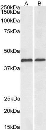
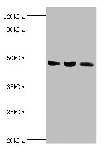
Anti-ACAT1 Antibody
HPA004428
ApplicationsWestern Blot, ImmunoCytoChemistry, ImmunoHistoChemistry
Product group Antibodies
ReactivityHuman, Mouse, Rat
TargetACAT1
Overview
- SupplierAtlas Antibodies
- Product NameAnti-ACAT1 Antibody
- Delivery Days Customer4
- ApplicationsWestern Blot, ImmunoCytoChemistry, ImmunoHistoChemistry
- CertificationResearch Use Only
- ClonalityPolyclonal
- ConjugateUnconjugated
- Gene ID38
- Target nameACAT1
- Target descriptionacetyl-CoA acetyltransferase 1
- Target synonymsACAT, MAT, T2, THIL, acetyl-CoA acetyltransferase, mitochondrial, acetoacetyl Coenzyme A thiolase, acetoacetyl-CoA thiolase, acetyl-Coenzyme A acetyltransferase 1, mitochondrial acetoacetyl-CoA thiolase, testicular tissue protein Li 198
- HostRabbit
- IsotypeIgG
- Protein IDP24752
- Protein NameAcetyl-CoA acetyltransferase, mitochondrial
- Scientific DescriptionRecombinant Protein Epitope Signature Tag (PrEST) antigen sequence
- ReactivityHuman, Mouse, Rat
- Storage Instruction-20°C,2°C to 8°C
- UNSPSC41116161
Datasheet
MSDS
Related products
Product group Antibodies
ApplicationsWestern Blot, ELISA
ReactivityHuman, Mouse, Rat
- SizePrice
Product group Antibodies
Anti-ACAT1 Antibody144-60258
ApplicationsWestern Blot, ImmunoHistoChemistry
ReactivityHuman, Mouse
TargetACAT1
- SizePrice
Product group Antibodies

ACAT1 AntibodyLS-C748331
ApplicationsWestern Blot, ImmunoHistoChemistry
ReactivityHuman, Mouse
TargetACAT1
- SizePrice

Product group Antibodies
Anti-ACAT1 Antibody Picoband(r)A02008-1-CARRIER-FREE
ApplicationsFlow Cytometry, ImmunoFluorescence, Western Blot, ELISA, ImmunoCytoChemistry, ImmunoHistoChemistry
ReactivityHuman, Mouse, Rat
TargetACAT1
- SizePrice
Product group Antibodies
ApplicationsFlow Cytometry, Western Blot, ImmunoCytoChemistry
ReactivityHuman, Rat
TargetACAT1
- SizePrice
Product group Antibodies
ACAT1 AntibodyCSB-PA001134ESR1HU
ApplicationsWestern Blot, ELISA, ImmunoHistoChemistry
ReactivityHuman, Mouse, Rat
TargetACAT1
- SizePrice
Product group Antibodies

ApplicationsImmunoFluorescence, Western Blot, ELISA
ReactivityBovine, Human, Mouse, Rat
TargetACAT1
- SizePrice
Product group Antibodies
ACAT1 Polyclonal AntibodyCAC14809
ApplicationsWestern Blot, ELISA, ImmunoHistoChemistry
ReactivityMouse, Rat
TargetACAT1
- SizePrice