

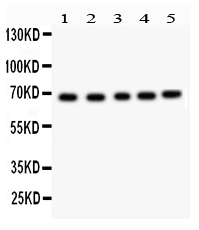
Figure 1. Western blot analysis of ACHE using anti-ACHE antibody (PB9417). Electrophoresis was performed on a 5-20% SDS-PAGE gel at 70V (Stacking gel) / 90V (Resolving gel) for 2-3 hours. Lane 1: Rat Kidney Tissue Lysate at 50ug, Lane 2: Mouse Liver Tissue Lysate at 50ug, Lane 3: HELA Whole Cell Lysate at 40ug, Lane 4: PANC Whole Cell Lysate at 40ug, Lane 5: COLO320 Whole Cell Lysate at 40ug. After electrophoresis, proteins were transferred to a nitrocellulose membrane at 150 mA for 50-90 minutes. Blocked the membrane with 5% non-fat milk/TBS for 1.5 hour at RT. The membrane was incubated with rabbit anti-ACHE antigen affinity purified polyclonal antibody (Catalog # PB9417) at 0.5 microg/mL overnight at 4°C, then washed with TBS-0.1%Tween 3 times with 5 minutes each and probed with a goat anti-rabbit IgG-HRP secondary antibody at a dilution of 1:5000 for 1.5 hour at RT. The signal is developed using an Enhanced Chemiluminescent detection (ECL) kit (Catalog # EK1002) with Tanon 5200 system. A specific band was detected for ACHE at approximately 68 kDa. The expected band size for ACHE is at 68 kDa.
Figure 1. Western blot analysis of ACHE using anti-ACHE antibody (PB9417). Electrophoresis was performed on a 5-20% SDS-PAGE gel at 70V (Stacking gel) / 90V (Resolving gel) for 2-3 hours. Lane 1: Rat Kidney Tissue Lysate at 50ug, Lane 2: Mouse Liver Tissue Lysate at 50ug, Lane 3: HELA Whole Cell Lysate at 40ug, Lane 4: PANC Whole Cell Lysate at 40ug, Lane 5: COLO320 Whole Cell Lysate at 40ug. After electrophoresis, proteins were transferred to a nitrocellulose membrane at 150 mA for 50-90 minutes. Blocked the membrane with 5% non-fat milk/TBS for 1.5 hour at RT. The membrane was incubated with rabbit anti-ACHE antigen affinity purified polyclonal antibody (Catalog # PB9417) at 0.5 microg/mL overnight at 4°C, then washed with TBS-0.1%Tween 3 times with 5 minutes each and probed with a goat anti-rabbit IgG-HRP secondary antibody at a dilution of 1:5000 for 1.5 hour at RT. The signal is developed using an Enhanced Chemiluminescent detection (ECL) kit (Catalog # EK1002) with Tanon 5200 system. A specific band was detected for ACHE at approximately 68 kDa. The expected band size for ACHE is at 68 kDa.
Anti-ACHE Picoband Antibody
PB9417
ApplicationsWestern Blot
Product group Antibodies
ReactivityHamster, Human, Mouse, Rat
TargetACHE
Overview
- SupplierBoster Bio
- Product NameAnti-ACHE Picoband Antibody
- Delivery Days Customer9
- Application Supplier NoteWB: The detection limit for ACHE is approximately 0.1ng/lane under reducing conditions. Tested Species: In-house tested species with positive results. Other applications have not been tested. Optimal dilutions should be determined by end users.
- ApplicationsWestern Blot
- CertificationResearch Use Only
- ClonalityPolyclonal
- Concentration500 ug/ml
- Gene ID43
- Target nameACHE
- Target descriptionacetylcholinesterase (Yt blood group)
- Target synonymsACEE, ARACHE, N-ACHE, YT, acetylcholinesterase, Yt blood group, acetylcholinesterase (Cartwright blood group), apoptosis-related acetylcholinesterase
- HostRabbit
- IsotypeIgG
- Protein IDP22303
- Protein NameAcetylcholinesterase
- Scientific DescriptionBoster Bio Anti-Acetylcholinesterase/ACHE Antibody Picoband® catalog # PB9417. Tested in WB applications. This antibody reacts with Human, Mouse, Rat. The brand Picoband indicates this is a premium antibody that guarantees superior quality, high affinity, and strong signals with minimal background in Western blot applications. Only our best-performing antibodies are designated as Picoband, ensuring unmatched performance.
- ReactivityHamster, Human, Mouse, Rat
- Storage Instruction-20°C,2°C to 8°C
- UNSPSC12352203
References
- Zhou Y, Dai Z, Deng K, et al. Eight Zhes Decoction ameliorates the lipid dysfunction of nonalcoholic fatty liver disease using integrated lipidomics, network pharmacology and pharmacokinetics. J Pharm Anal. 2023,13(9):1058-1069. doi: 10.1016/j.jpha.2023.05.012Read this paper
- Cao J, Wang K, Li N, et al. Soluble dietary fiber and cellulose from Saccharina japonica by-product ameliorate Loperamide-induced constipation via modulating enteric neurotransmitters, short-chain fatty acids and gut microbiota. Int J Biol Macromol. 2023,226:1319-1331. doi: 10.1016/j.ijbiomac.2022.11.243Read this paper
Datasheet
MSDS
Related products
Product group Antibodies
References
ACHE Polyclonal AntibodyBS-2511R
ApplicationsImmunoFluorescence, Western Blot, ELISA, ImmunoCytoChemistry, ImmunoHistoChemistry, ImmunoHistoChemistry Frozen, ImmunoHistoChemistry Paraffin
ReactivityBovine, Canine, Equine, Human, Mouse, Rat
TargetACHE
- SizePrice
Product group Antibodies
ApplicationsWestern Blot, ELISA, ImmunoCytoChemistry, ImmunoHistoChemistry, ImmunoHistoChemistry Frozen, ImmunoHistoChemistry Paraffin
ReactivityMouse, Rat
TargetACHE
- SizePrice
Product group Antibodies
Anti-ACHE Antibody144-02806
ApplicationsWestern Blot, ImmunoHistoChemistry
ReactivityHuman, Mouse, Rat
TargetACHE
- SizePrice
Product group Antibodies

Anti-acetylcholinesterase [1G]AB04029-1.1
ApplicationsELISA
ReactivityHuman
TargetACHE
- SizePrice
Product group Antibodies
Anti-AChE AntibodyA96247
ApplicationsWestern Blot, ELISA, ImmunoHistoChemistry
ReactivityHuman, Mouse, Rat
- SizePrice
Product group Antibodies

Goat anti-ACHE AntibodyEB06659
ApplicationsFlow Cytometry, ImmunoFluorescence, Western Blot, ELISA
ReactivityHuman, Mouse, Rat
TargetACHE
- SizePrice
Product group Antibodies
References
AChE antibodyGTX101648
ApplicationsImmunoFluorescence, Western Blot, ImmunoCytoChemistry, ImmunoHistoChemistry, ImmunoHistoChemistry Frozen, ImmunoHistoChemistry Paraffin
ReactivityHuman, Mouse, Rat
TargetACHE
- SizePrice