


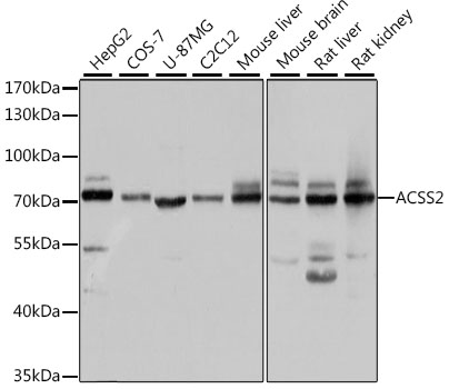
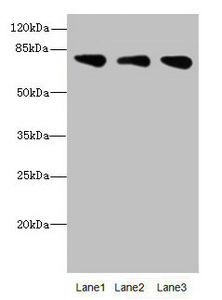
Figure 1. Western blot analysis of ACSS2 using anti-ACSS2 antibody (A02809-1). Electrophoresis was performed on a 5-20% SDS-PAGE gel at 70V (Stacking gel) / 90V (Resolving gel) for 2-3 hours. The sample well of each lane was loaded with 30 ug of sample under reducing conditions. Lane 1: human HepG2 whole cell lysates, Lane 2: human Hela whole cell lysates, Lane 3: human U-87MG whole cell lysates, Lane 4: human MCF-7 whole cell lysates, Lane 5: rat liver tissue lysates, Lane 6: rat C6 whole cell lysates, Lane 7: mouse liver tissue lysates. After electrophoresis, proteins were transferred to a nitrocellulose membrane at 150 mA for 50-90 minutes. Blocked the membrane with 5% non-fat milk/TBS for 1.5 hour at RT. The membrane was incubated with rabbit anti-ACSS2 antigen affinity purified polyclonal antibody (Catalog # A02809-1) at 0.25 microg/mL overnight at 4°C, then washed with TBS-0.1%Tween 3 times with 5 minutes each and probed with a goat anti-rabbit IgG-HRP secondary antibody at a dilution of 1:5000 for 1.5 hour at RT. The signal is developed using an Enhanced Chemiluminescent detection (ECL) kit (Catalog # EK1002) with Tanon 5200 system. A specific band was detected for ACSS2 at approximately 79 kDa. The expected band size for ACSS2 is at 79 kDa.
. Overlay histogram showing U20S cells stained with A02809-1 (Blue line). To facilitate intracellular staining, cells were fixed with 4% paraformaldehyde and permeabilized with permeabilization buffer. The cells were blocked with 10% normal goat serum. And then incubated with rabbit anti-ACSS2 Antibody (A02809-1, 1 microg/1x106 cells) for 30 min at 20°C. DyLight®488 conjugated goat anti-rabbit IgG (BA1127, 5-10 microg/1x106 cells) was used as secondary antibody for 30 minutes at 20°C. Isotype control antibody (Green line) was rabbit IgG (1 microg/1x106) used under the same conditions. Unlabelled sample without incubation with primary antibody and secondary antibody (Red line) was used as a blank control.")
Figure 1. Western blot analysis of ACSS2 using anti-ACSS2 antibody (A02809-1). Electrophoresis was performed on a 5-20% SDS-PAGE gel at 70V (Stacking gel) / 90V (Resolving gel) for 2-3 hours. The sample well of each lane was loaded with 30 ug of sample under reducing conditions. Lane 1: human HepG2 whole cell lysates, Lane 2: human Hela whole cell lysates, Lane 3: human U-87MG whole cell lysates, Lane 4: human MCF-7 whole cell lysates, Lane 5: rat liver tissue lysates, Lane 6: rat C6 whole cell lysates, Lane 7: mouse liver tissue lysates. After electrophoresis, proteins were transferred to a nitrocellulose membrane at 150 mA for 50-90 minutes. Blocked the membrane with 5% non-fat milk/TBS for 1.5 hour at RT. The membrane was incubated with rabbit anti-ACSS2 antigen affinity purified polyclonal antibody (Catalog # A02809-1) at 0.25 microg/mL overnight at 4°C, then washed with TBS-0.1%Tween 3 times with 5 minutes each and probed with a goat anti-rabbit IgG-HRP secondary antibody at a dilution of 1:5000 for 1.5 hour at RT. The signal is developed using an Enhanced Chemiluminescent detection (ECL) kit (Catalog # EK1002) with Tanon 5200 system. A specific band was detected for ACSS2 at approximately 79 kDa. The expected band size for ACSS2 is at 79 kDa.
Anti-ACSS2 Antibody Picoband(r)
A02809-1-CARRIER-FREE
ApplicationsFlow Cytometry, Western Blot, ELISA
Product group Antibodies
ReactivityHuman, Mouse, Rat
TargetACSS2
Overview
- SupplierBoster Bio
- Product NameAnti-ACSS2 Antibody Picoband(r)
- Delivery Days Customer9
- ApplicationsFlow Cytometry, Western Blot, ELISA
- CertificationResearch Use Only
- ClonalityPolyclonal
- Concentration500 ug/ml
- Gene ID55902
- Target nameACSS2
- Target descriptionacyl-CoA synthetase short chain family member 2
- Target synonymsACAS2, ACECS, ACS, ACSA, AceCS1, dJ1161H23.1, acetyl-coenzyme A synthetase, cytoplasmic, acetate thiokinase, acetate-CoA ligase, acetyl-CoA synthetase 1, acetyl-Coenzyme A synthetase 2 (ADP forming), acyl-activating enzyme, cytoplasmic acetyl-coenzyme A synthetase, propionate--CoA ligase
- HostRabbit
- IsotypeIgG
- Protein IDQ9NR19
- Protein NameAcetyl-coenzyme A synthetase, cytoplasmic
- Scientific DescriptionBoster Bio Anti-ACSS2 Antibody Picoband® catalog # A02809-1. Tested in ELISA, Flow Cytometry, WB applications. This antibody reacts with Human, Mouse, Rat. The brand Picoband indicates this is a premium antibody that guarantees superior quality, high affinity, and strong signals with minimal background in Western blot applications. Only our best-performing antibodies are designated as Picoband, ensuring unmatched performance.
- ReactivityHuman, Mouse, Rat
- Storage Instruction-20°C,2°C to 8°C
- UNSPSC12352203
Related products
Product group Antibodies
Anti-ACSS2 AntibodyA15241
ApplicationsImmunoFluorescence, Western Blot, ImmunoCytoChemistry
ReactivityHuman, Mouse, Rat
- SizePrice
Product group Antibodies
Anti-ACSS2 Antibody144-66151
ApplicationsImmunoFluorescence, Western Blot, ImmunoHistoChemistry
ReactivityHuman, Mouse, Rat
TargetACSS2
- SizePrice
Product group Antibodies

ACSS2 / ACAS2 AntibodyLS-C748639
ApplicationsImmunoFluorescence, Western Blot, ImmunoHistoChemistry
ReactivityHuman, Mouse, Rat
TargetACSS2
- SizePrice
Product group Antibodies
ACSS2 Recombinant Antibody, Biotin ConjugatedBSM-61364R-BIOTIN
ApplicationsWestern Blot
ReactivityHuman, Mouse, Rat
TargetACSS2
- SizePrice
Product group Antibodies
ACSS2 AntibodyCSB-PA873629ESR2HU
ApplicationsWestern Blot, ELISA, ImmunoHistoChemistry
ReactivityHuman, Mouse
TargetACSS2
- SizePrice
Product group Antibodies
Acss2 Polyclonal AntibodyCAC10705
ApplicationsWestern Blot, ELISA, ImmunoHistoChemistry
ReactivityMouse
TargetACSS2
- SizePrice
Product group Antibodies
References
ACSS2 antibodyGTX30020
ApplicationsImmunoFluorescence, Western Blot, ImmunoCytoChemistry, ImmunoHistoChemistry, ImmunoHistoChemistry Paraffin
ReactivityHuman, Monkey, Mouse, Rat
TargetACSS2
- SizePrice

Product group Antibodies
Anti-ACSS2 AntibodyHPA004141
ApplicationsWestern Blot, ImmunoCytoChemistry, ImmunoHistoChemistry
ReactivityHuman
TargetACSS2
- SizePrice
Product group Antibodies
Anti-ACSS2 AntibodyCAB6472
ApplicationsImmunoFluorescence, Western Blot, ELISA, ImmunoCytoChemistry
ReactivityHuman
TargetACSS2
- SizePrice