


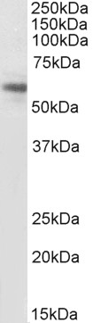
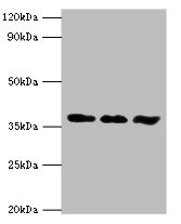
Figure 1. Western blot analysis of ACVR1 using anti-ACVR1 antibody (M00798). Electrophoresis was performed on a 5-20% SDS-PAGE gel at 70V (Stacking gel) / 90V (Resolving gel) for 2-3 hours. The sample well of each lane was loaded with 30 ug of sample under reducing conditions. Lane 1: rat brain tissue lysates, Lane 2: rat heart tissue lysates, Lane 3: mouse brain tissue lysates, Lane 4: mouse heart tissue lysates. After electrophoresis, proteins were transferred to a nitrocellulose membrane at 150 mA for 50-90 minutes. Blocked the membrane with 5% non-fat milk/TBS for 1.5 hour at RT. The membrane was incubated with rabbit anti-ACVR1 antigen affinity purified monoclonal antibody (Catalog # M00798) at 1:500 overnight at 4°C, then washed with TBS-0.1%Tween 3 times with 5 minutes each and probed with a goat anti-rabbit IgG-HRP secondary antibody at a dilution of 1:5000 for 1.5 hour at RT. The signal is developed using an Enhanced Chemiluminescent detection (ECL) kit (Catalog # EK1002) with Tanon 5200 system. A specific band was detected for ACVR1 at approximately 57 kDa. The expected band size for ACVR1 is at 57 kDa.
Figure 1. Western blot analysis of ACVR1 using anti-ACVR1 antibody (M00798). Electrophoresis was performed on a 5-20% SDS-PAGE gel at 70V (Stacking gel) / 90V (Resolving gel) for 2-3 hours. The sample well of each lane was loaded with 30 ug of sample under reducing conditions. Lane 1: rat brain tissue lysates, Lane 2: rat heart tissue lysates, Lane 3: mouse brain tissue lysates, Lane 4: mouse heart tissue lysates. After electrophoresis, proteins were transferred to a nitrocellulose membrane at 150 mA for 50-90 minutes. Blocked the membrane with 5% non-fat milk/TBS for 1.5 hour at RT. The membrane was incubated with rabbit anti-ACVR1 antigen affinity purified monoclonal antibody (Catalog # M00798) at 1:500 overnight at 4°C, then washed with TBS-0.1%Tween 3 times with 5 minutes each and probed with a goat anti-rabbit IgG-HRP secondary antibody at a dilution of 1:5000 for 1.5 hour at RT. The signal is developed using an Enhanced Chemiluminescent detection (ECL) kit (Catalog # EK1002) with Tanon 5200 system. A specific band was detected for ACVR1 at approximately 57 kDa. The expected band size for ACVR1 is at 57 kDa.
Anti-ACVR1 Rabbit Monoclonal Antibody
M00798
ApplicationsImmunoPrecipitation, Western Blot
Product group Antibodies
ReactivityHuman, Mouse, Rat
TargetACVR1
Overview
- SupplierBoster Bio
- Product NameAnti-ACVR1 Rabbit Monoclonal Antibody
- Delivery Days Customer9
- ApplicationsImmunoPrecipitation, Western Blot
- CertificationResearch Use Only
- ClonalityMonoclonal
- Clone ID25A99
- Gene ID90
- Target nameACVR1
- Target descriptionactivin A receptor type 1
- Target synonymsACTRI, ACVR1A, ACVRLK2, ALK2, FOP, SKR1, TSRI, activin receptor type-1, TGF-B superfamily receptor type I, activin A receptor, type I, activin A receptor, type II-like kinase 2, activin receptor type I, activin receptor-like kinase 2, hydroxyalkyl-protein kinase, serine/threonine-protein kinase receptor R1
- HostRabbit
- IsotypeIgG
- Protein IDQ04771
- Protein NameActivin receptor type-1
- Scientific DescriptionBoster Bio Anti-ACVR1 Rabbit Monoclonal Antibody catalog # M00798. Tested in WB, IP applications. This antibody reacts with Human, Mouse, Rat.
- ReactivityHuman, Mouse, Rat
- Storage Instruction-20°C
- UNSPSC12352203
References
- Wang J, You H, Qi J, et al. Autocrine and paracrine STIP1 signaling promote osteolytic bone metastasis in renal cell carcinoma. Oncotarget. 2017,8(10):17012-17026. doi: 10.18632/oncotarget.15222Read this paper
Related products
Product group Antibodies
ApplicationsWestern Blot, ELISA
ReactivityHuman
- SizePrice
Product group Antibodies
Anti-ACVR1 Antibody101-10021
ApplicationsWestern Blot, ELISA
TargetACVR1
- SizePrice
Product group Antibodies
ApplicationsImmunoPrecipitation, Western Blot
ReactivityHuman, Mouse, Rat
TargetACVR1
- SizePrice
Product group Antibodies
ACVR1 AntibodyCSB-PA001257ESR2HU
ApplicationsImmunoFluorescence, Western Blot, ELISA, ImmunoHistoChemistry
ReactivityHuman, Mouse
TargetACVR1
- SizePrice
Product group Antibodies

Goat anti-ACVR1, BiotinylatedEB08207-B
ApplicationsWestern Blot, ELISA, ImmunoHistoChemistry
ReactivityCanine, Human, Mouse, Rat
TargetACVR1
- SizePrice
Product group Antibodies
ApplicationsImmunoPrecipitation, Western Blot, ImmunoCytoChemistry, ImmunoHistoChemistry
ReactivityMouse, Porcine, Rat
TargetACVR1
- SizePrice

Product group Antibodies
Anti-ACVR1 AntibodyHPA007505
ApplicationsImmunoHistoChemistry
ReactivityHuman
TargetACVR1
- SizePrice
Product group Antibodies
Activin Receptor Type IA antibodyGTX107815
ApplicationsWestern Blot, ImmunoHistoChemistry, ImmunoHistoChemistry Frozen
ReactivityHuman, Mouse, Rat
TargetACVR1
- SizePrice
Product group Antibodies
TargetACVR1
- SizePrice
Product group Antibodies

ALK2 / ACVR1 AntibodyLS-C404684
ApplicationsELISA, ImmunoHistoChemistry
ReactivityHuman, Mouse, Rat
TargetACVR1
- SizePrice