


Anti-ACVRL1 Antibody
A96975
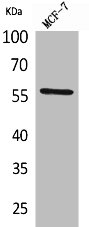
ApplicationsWestern Blot, ELISA
Product group Antibodies
ReactivityHuman, Mouse, Rat
Overview
- SupplierAntibodies.com
- Product NameAnti-ACVRL1 Antibody
- Delivery Days Customer7
- ApplicationsWestern Blot, ELISA
- CertificationResearch Use Only
- ClonalityPolyclonal
- ConjugateUnconjugated
- HostRabbit
- IsotypeIgG
- Scientific DescriptionRabbit polyclonal antibody to ACVRL1.
- ReactivityHuman, Mouse, Rat
- UNSPSC12352203
Related products
Product group Antibodies
ReactivityHuman
TargetACVRL1
- SizePrice
Product group Antibodies
ACVRL1 AntibodyCSB-PA005080
ApplicationsWestern Blot, ELISA
ReactivityHuman, Mouse, Rat
TargetACVRL1
- SizePrice

Product group Antibodies
Anti-ALK-1/ACVRL1 Antibody Picoband(r)A01468-2-CARRIER-FREE
ApplicationsFlow Cytometry, Western Blot, ELISA, ImmunoHistoChemistry
ReactivityHuman, Mouse, Rat
TargetACVRL1
- SizePrice
Product group Antibodies

ApplicationsELISA
ReactivityHuman
TargetACVRL1
- SizePrice
Product group Antibodies

ApplicationsWestern Blot, ELISA
ReactivityHuman
TargetACVRL1
- SizePrice

Product group Antibodies
Anti-ACVRL1 AntibodyHPA007041
ApplicationsWestern Blot, ImmunoHistoChemistry
ReactivityHuman
TargetACVRL1
- SizePrice
Product group Antibodies
ACVRL1 Polyclonal AntibodyCAC14534
ApplicationsImmunoFluorescence, Western Blot, ELISA
TargetACVRL1
- SizePrice
Product group Antibodies
ACVRL1 antibody [C3], C-termGTX100035
ApplicationsWestern Blot, ImmunoHistoChemistry, ImmunoHistoChemistry Paraffin
ReactivityHuman, Mouse
TargetACVRL1
- SizePrice
Product group Antibodies
Anti-ACVRL1Y058240
ApplicationsWestern Blot, ELISA, ImmunoHistoChemistry
ReactivityHuman
- SizePrice
Product group Antibodies
ACVRL1 Recombinant Antibody, AbBy Fluor-594 ConjugatedBSM-61519R-BF594
ApplicationsWestern Blot
ReactivityHuman, Mouse, Rat
TargetACVRL1
- SizePrice