


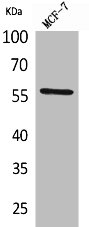
Figure 1. Western blot analysis of ALK-1/ACVRL1 using anti-ALK-1/ACVRL1 antibody (A01468-2). Electrophoresis was performed on a 5-20% SDS-PAGE gel at 70V (Stacking gel) / 90V (Resolving gel) for 2-3 hours. The sample well of each lane was loaded with 30ug of sample under reducing conditions. Lane 1: human HL-60 whole cell lysates, Lane 2: human THP-1 whole cell lysates, Lane 3: rat brain tissue lysates, Lane 4: rat heart tissue lysates, Lane 5: mouse brain tissue lysates, Lane 6: mouse kidney tissue lysates, Lane 7: mouse heart tissue lysates. After Electrophoresis, proteins were transferred to a Nitrocellulose membrane at 150mA for 50-90 minutes. Blocked the membrane with 5% Non-fat Milk/ TBS for 1.5 hour at RT. The membrane was incubated with rabbit anti-ALK-1/ACVRL1 antigen affinity purified polyclonal antibody (Catalog # A01468-2) at 0.5 microg/mL overnight at 4°C, then washed with TBS-0.1%Tween 3 times with 5 minutes each and probed with a goat anti-rabbit IgG-HRP secondary antibody at a dilution of 1:5000 for 1.5 hour at RT. The signal is developed using an Enhanced Chemiluminescent detection (ECL) kit (Catalog # EK1002) with Tanon 5200 system. A specific band was detected for ALK-1/ACVRL1 at approximately 55KD. The expected band size for ALK-1/ACVRL1 is at 55KD.
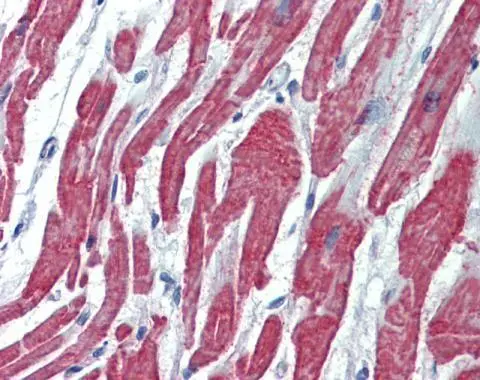
. ALK-1/ACVRL1 was detected in paraffin-embedded section of human breast cancer tissue. Heat mediated antigen retrieval was performed in EDTA buffer (pH8.0, epitope retrieval solution). The tissue section was blocked with 10% goat serum. The tissue section was then incubated with 2microg/ml rabbit anti-ALK-1/ACVRL1 Antibody (A01468-2) overnight at 4°C. Biotinylated goat anti-rabbit IgG was used as secondary antibody and incubated for 30 minutes at 37°C. The tissue section was developed using Strepavidin-Biotin-Complex (SABC) (Catalog # SA1022) with DAB as the chromogen.")
. ALK-1/ACVRL1 was detected in paraffin-embedded section of human gallbladder adenocarcinoma tissue. Heat mediated antigen retrieval was performed in EDTA buffer (pH8.0, epitope retrieval solution). The tissue section was blocked with 10% goat serum. The tissue section was then incubated with 2microg/ml rabbit anti-ALK-1/ACVRL1 Antibody (A01468-2) overnight at 4°C. Biotinylated goat anti-rabbit IgG was used as secondary antibody and incubated for 30 minutes at 37°C. The tissue section was developed using Strepavidin-Biotin-Complex (SABC) (Catalog # SA1022) with DAB as the chromogen.")
. ALK-1/ACVRL1 was detected in paraffin-embedded section of human liver cancer tissue. Heat mediated antigen retrieval was performed in EDTA buffer (pH8.0, epitope retrieval solution). The tissue section was blocked with 10% goat serum. The tissue section was then incubated with 2microg/ml rabbit anti-ALK-1/ACVRL1 Antibody (A01468-2) overnight at 4°C. Biotinylated goat anti-rabbit IgG was used as secondary antibody and incubated for 30 minutes at 37°C. The tissue section was developed using Strepavidin-Biotin-Complex (SABC) (Catalog # SA1022) with DAB as the chromogen.")
. ALK-1/ACVRL1 was detected in paraffin-embedded section of human ovarian serous adenocarcinoma tissue. Heat mediated antigen retrieval was performed in EDTA buffer (pH8.0, epitope retrieval solution). The tissue section was blocked with 10% goat serum. The tissue section was then incubated with 2microg/ml rabbit anti-ALK-1/ACVRL1 Antibody (A01468-2) overnight at 4°C. Biotinylated goat anti-rabbit IgG was used as secondary antibody and incubated for 30 minutes at 37°C. The tissue section was developed using Strepavidin-Biotin-Complex (SABC) (Catalog # SA1022) with DAB as the chromogen.")
. Overlay histogram showing MCF-7 cells stained with A01468-2 (Blue line). The cells were fixed with 4% paraformaldehyde and blocked with 10% normal goat serum. And then incubated with rabbit anti-ALK-1/ACVRL1 Antibody (A01468-2, 1microg/1x106 cells) for 30 min at 20°C. DyLight®488 conjugated goat anti-rabbit IgG (BA1127, 5-10microg/1x106 cells) was used as secondary antibody for 30 minutes at 20°C. Isotype control antibody (Green line) was rabbit IgG (1microg/1x106) used under the same conditions. Unlabelled sample without incubation with primary antibody and secondary antibody (Red line) was used as a blank control.")
Figure 1. Western blot analysis of ALK-1/ACVRL1 using anti-ALK-1/ACVRL1 antibody (A01468-2). Electrophoresis was performed on a 5-20% SDS-PAGE gel at 70V (Stacking gel) / 90V (Resolving gel) for 2-3 hours. The sample well of each lane was loaded with 30ug of sample under reducing conditions. Lane 1: human HL-60 whole cell lysates, Lane 2: human THP-1 whole cell lysates, Lane 3: rat brain tissue lysates, Lane 4: rat heart tissue lysates, Lane 5: mouse brain tissue lysates, Lane 6: mouse kidney tissue lysates, Lane 7: mouse heart tissue lysates. After Electrophoresis, proteins were transferred to a Nitrocellulose membrane at 150mA for 50-90 minutes. Blocked the membrane with 5% Non-fat Milk/ TBS for 1.5 hour at RT. The membrane was incubated with rabbit anti-ALK-1/ACVRL1 antigen affinity purified polyclonal antibody (Catalog # A01468-2) at 0.5 microg/mL overnight at 4°C, then washed with TBS-0.1%Tween 3 times with 5 minutes each and probed with a goat anti-rabbit IgG-HRP secondary antibody at a dilution of 1:5000 for 1.5 hour at RT. The signal is developed using an Enhanced Chemiluminescent detection (ECL) kit (Catalog # EK1002) with Tanon 5200 system. A specific band was detected for ALK-1/ACVRL1 at approximately 55KD. The expected band size for ALK-1/ACVRL1 is at 55KD.
Anti-ALK-1/ACVRL1 Antibody Picoband(r)
A01468-2-CARRIER-FREE
ApplicationsFlow Cytometry, Western Blot, ELISA, ImmunoHistoChemistry
Product group Antibodies
ReactivityHuman, Mouse, Rat
TargetACVRL1
Overview
- SupplierBoster Bio
- Product NameAnti-ALK-1/ACVRL1 Antibody Picoband(r)
- Delivery Days Customer9
- ApplicationsFlow Cytometry, Western Blot, ELISA, ImmunoHistoChemistry
- CertificationResearch Use Only
- ClonalityPolyclonal
- Concentration500 ug/ml
- Gene ID94
- Target nameACVRL1
- Target descriptionactivin A receptor like type 1
- Target synonymsACVRLK1, ALK-1, ALK1, HHT, HHT2, ORW2, SKR3, TSR-I, activin receptor type-1-like, TGF-B superfamily receptor type I, activin A receptor type II-like 1, activin A receptor type IL, activin A receptor, type II-like kinase 1, serine/threonine-protein kinase receptor R3
- HostRabbit
- IsotypeIgG
- Protein IDP37023
- Protein NameActivin receptor type-1-like
- Scientific DescriptionBoster Bio Anti-ALK-1/ACVRL1 Antibody Picoband® catalog # A01468-2. Tested in ELISA, Flow Cytometry, IHC, WB applications. This antibody reacts with Human, Mouse, Rat. The brand Picoband indicates this is a premium antibody that guarantees superior quality, high affinity, and strong signals with minimal background in Western blot applications. Only our best-performing antibodies are designated as Picoband, ensuring unmatched performance.
- ReactivityHuman, Mouse, Rat
- Storage Instruction-20°C,2°C to 8°C
- UNSPSC12352203
Related products
Product group Antibodies
ReactivityHuman
TargetACVRL1
- SizePrice
Product group Antibodies
ACVRL1 AntibodyCSB-PA005080
ApplicationsWestern Blot, ELISA
ReactivityHuman, Mouse, Rat
TargetACVRL1
- SizePrice

Product group Antibodies
Anti-ACVRL1 AntibodyA96975
ApplicationsWestern Blot, ELISA
ReactivityHuman, Mouse, Rat
- SizePrice
Product group Antibodies

ApplicationsELISA
ReactivityHuman
TargetACVRL1
- SizePrice
Product group Antibodies

ApplicationsWestern Blot, ELISA
ReactivityHuman
TargetACVRL1
- SizePrice

Product group Antibodies
Anti-ACVRL1 AntibodyHPA007041
ApplicationsWestern Blot, ImmunoHistoChemistry
ReactivityHuman
TargetACVRL1
- SizePrice
Product group Antibodies
ACVRL1 Polyclonal AntibodyCAC14534
ApplicationsImmunoFluorescence, Western Blot, ELISA
TargetACVRL1
- SizePrice
Product group Antibodies
ACVRL1 antibody [C3], C-termGTX100035
ApplicationsWestern Blot, ImmunoHistoChemistry, ImmunoHistoChemistry Paraffin
ReactivityHuman, Mouse
TargetACVRL1
- SizePrice
Product group Antibodies
Anti-ACVRL1Y058240
ApplicationsWestern Blot, ELISA, ImmunoHistoChemistry
ReactivityHuman
- SizePrice
Product group Antibodies
ACVRL1 Recombinant Antibody, AbBy Fluor-594 ConjugatedBSM-61519R-BF594
ApplicationsWestern Blot
ReactivityHuman, Mouse, Rat
TargetACVRL1
- SizePrice