


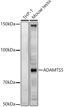
Figure 1. Western blot analysis of ADAMTS5 using anti-ADAMTS5 antibody (A02802-1). Electrophoresis was performed on a 5-20% SDS-PAGE gel at 70V (Stacking gel) / 90V (Resolving gel) for 2-3 hours. The sample well of each lane was loaded with 30 ug of sample under reducing conditions. Lane 1: human Hela whole cell lysates, Lane 2: human HepG2 whole cell lysates, Lane 3: human U20S whole cell lysates, Lane 4: human SW620 whole cell lysates, Lane 5: human THP-1 whole cell lysates, Lane 6: human Raji whole cell lysates, Lane 7: human SGC-7901 whole cell lysates, Lane 8: rat liver tissue lysates, Lane 9: rat testis tissue lysates, Lane 10: mouse liver tissue lysates, Lane 11: mouse testis tissue lysates, After electrophoresis, proteins were transferred to a nitrocellulose membrane at 150 mA for 50-90 minutes. Blocked the membrane with 5% non-fat milk/TBS for 1.5 hour at RT. The membrane was incubated with rabbit anti-ADAMTS5 antigen affinity purified polyclonal antibody (Catalog # A02802-1) at 0.5 microg/mL overnight at 4°C, then washed with TBS-0.1%Tween 3 times with 5 minutes each and probed with a goat anti-rabbit IgG-HRP secondary antibody at a dilution of 1:5000 for 1.5 hour at RT. The signal is developed using an Enhanced Chemiluminescent detection (ECL) kit (Catalog # EK1002) with Tanon 5200 system. A specific band was detected for ADAMTS5 at approximately 75 kDa. The expected band size for ADAMTS5 is at 102 kDa.
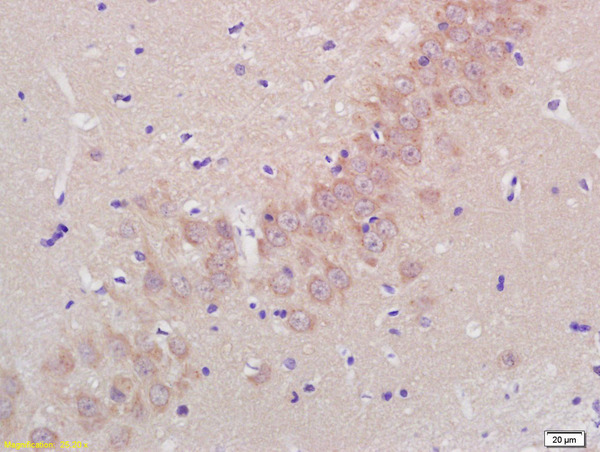
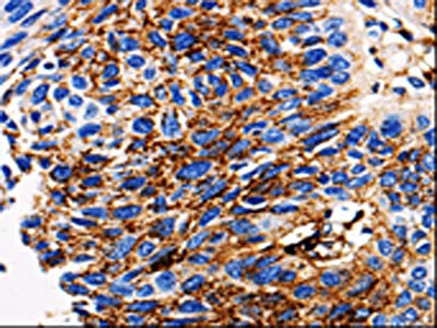
. ADAMTS5 was detected in a paraffin-embedded section of human placenta tissue. Heat mediated antigen retrieval was performed in EDTA buffer (pH 8.0, epitope retrieval solution). The tissue section was blocked with 10% goat serum. The tissue section was then incubated with 2 microg/ml rabbit anti-ADAMTS5 Antibody (A02802-1) overnight at 4°C. Biotinylated goat anti-rabbit IgG was used as secondary antibody and incubated for 30 minutes at 37°C. The tissue section was developed using Strepavidin-Biotin-Complex (SABC) (Catalog # SA1022) with DAB as the chromogen.")
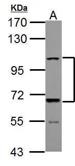
Figure 1. Western blot analysis of ADAMTS5 using anti-ADAMTS5 antibody (A02802-1). Electrophoresis was performed on a 5-20% SDS-PAGE gel at 70V (Stacking gel) / 90V (Resolving gel) for 2-3 hours. The sample well of each lane was loaded with 30 ug of sample under reducing conditions. Lane 1: human Hela whole cell lysates, Lane 2: human HepG2 whole cell lysates, Lane 3: human U20S whole cell lysates, Lane 4: human SW620 whole cell lysates, Lane 5: human THP-1 whole cell lysates, Lane 6: human Raji whole cell lysates, Lane 7: human SGC-7901 whole cell lysates, Lane 8: rat liver tissue lysates, Lane 9: rat testis tissue lysates, Lane 10: mouse liver tissue lysates, Lane 11: mouse testis tissue lysates, After electrophoresis, proteins were transferred to a nitrocellulose membrane at 150 mA for 50-90 minutes. Blocked the membrane with 5% non-fat milk/TBS for 1.5 hour at RT. The membrane was incubated with rabbit anti-ADAMTS5 antigen affinity purified polyclonal antibody (Catalog # A02802-1) at 0.5 microg/mL overnight at 4°C, then washed with TBS-0.1%Tween 3 times with 5 minutes each and probed with a goat anti-rabbit IgG-HRP secondary antibody at a dilution of 1:5000 for 1.5 hour at RT. The signal is developed using an Enhanced Chemiluminescent detection (ECL) kit (Catalog # EK1002) with Tanon 5200 system. A specific band was detected for ADAMTS5 at approximately 75 kDa. The expected band size for ADAMTS5 is at 102 kDa.
Anti-ADAMTS5 Antibody Picoband(r)
A02802-1-CARRIER-FREE
ApplicationsWestern Blot, ELISA, ImmunoHistoChemistry
Product group Antibodies
ReactivityHuman, Mouse, Rat
TargetADAMTS5
Overview
- SupplierBoster Bio
- Product NameAnti-ADAMTS5 Antibody Picoband(r)
- Delivery Days Customer9
- ApplicationsWestern Blot, ELISA, ImmunoHistoChemistry
- CertificationResearch Use Only
- ClonalityPolyclonal
- Concentration500 ug/ml
- Gene ID11096
- Target nameADAMTS5
- Target descriptionADAM metallopeptidase with thrombospondin type 1 motif 5
- Target synonymsADAM-TS 11, ADAM-TS 5, ADAM-TS5, ADAMTS-11, ADAMTS-5, ADAMTS11, ADMP-2, A disintegrin and metalloproteinase with thrombospondin motifs 5, a disintegrin and metalloproteinase with thrombospondin motifs 11, a disintegrin-like and metalloprotease (reprolysin type) with thrombospondin type 1 motif, 5 (aggrecanase-2), aggrecanase-2
- HostRabbit
- IsotypeIgG
- Protein IDQ9UNA0
- Protein NameA disintegrin and metalloproteinase with thrombospondin motifs 5
- Scientific DescriptionBoster Bio Anti-ADAMTS5 Antibody Picoband® catalog # A02802-1. Tested in ELISA, IHC, WB applications. This antibody reacts with Human, Mouse, Rat. The brand Picoband indicates this is a premium antibody that guarantees superior quality, high affinity, and strong signals with minimal background in Western blot applications. Only our best-performing antibodies are designated as Picoband, ensuring unmatched performance.
- ReactivityHuman, Mouse, Rat
- Storage Instruction-20°C,2°C to 8°C
- UNSPSC12352203
Related products
Product group Antibodies

Anti-ADAMTS5 [7B4]Ab03226-1.1
ApplicationsImmunoHistoChemistry, Neutralisation/Blocking, Other Application
ReactivityHuman, Mouse
TargetADAMTS5
- SizePrice
Product group Antibodies
Anti-ADAMTS5 Antibody144-02836
ApplicationsWestern Blot
ReactivityHuman, Mouse
TargetADAMTS5
- SizePrice
Product group Antibodies
Anti-ADAMTS5 AntibodyA14217
ApplicationsImmunoFluorescence, Western Blot, ImmunoCytoChemistry
ReactivityHuman, Mouse, Rat
- SizePrice
Product group Antibodies
References
ADAMTS5 Polyclonal AntibodyBS-3573R
ApplicationsImmunoFluorescence, Western Blot, ELISA, ImmunoHistoChemistry, ImmunoHistoChemistry Frozen, ImmunoHistoChemistry Paraffin
ReactivityBovine, Canine, Equine, Human, Mouse, Porcine, Rabbit, Rat
TargetADAMTS5
- SizePrice
Product group Antibodies
ADAMTS5 AntibodyCSB-PA198249
ApplicationsELISA, ImmunoHistoChemistry
ReactivityHuman, Mouse, Rat
TargetADAMTS5
- SizePrice
Product group Antibodies
ApplicationsImmunoPrecipitation, Western Blot, ImmunoCytoChemistry, ImmunoHistoChemistry
TargetADAMTS5
- SizePrice
Product group Antibodies

ADAMTS5 AntibodyLS-C408561
ApplicationsWestern Blot
ReactivityHuman, Mouse
TargetADAMTS5
- SizePrice
Product group Antibodies
ADAMTS5 antibodyGTX100332
ApplicationsWestern Blot, ImmunoHistoChemistry, ImmunoHistoChemistry Paraffin
ReactivityHuman, Mouse
TargetADAMTS5
- SizePrice

Product group Antibodies
Anti-ADAMTS5 AntibodyHPA005661
ApplicationsImmunoHistoChemistry
ReactivityHuman
TargetADAMTS5
- SizePrice