

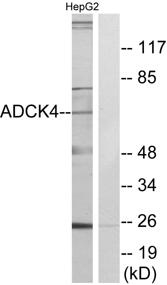


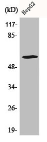
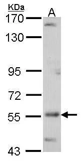
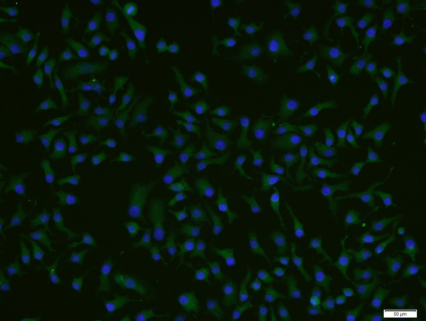
Anti-ADCK4 Antibody
A96093
ApplicationsImmunoFluorescence, Western Blot, ELISA
Product group Antibodies
ReactivityHuman, Mouse, Rat
Overview
- SupplierAntibodies.com
- Product NameAnti-ADCK4 Antibody
- Delivery Days Customer7
- ApplicationsImmunoFluorescence, Western Blot, ELISA
- CertificationResearch Use Only
- ClonalityPolyclonal
- ConjugateUnconjugated
- HostRabbit
- IsotypeIgG
- Scientific DescriptionRabbit polyclonal antibody to ADCK4.
- ReactivityHuman, Mouse, Rat
- UNSPSC12352203
Related products
Product group Antibodies
ADCK4 AntibodyCSB-PA000823
ApplicationsImmunoFluorescence, Western Blot, ELISA, ImmunoHistoChemistry
ReactivityHuman, Mouse, Rat
TargetCOQ8B
- SizePrice

Product group Antibodies
Anti-COQ8B Antibody Picoband(r)A32263-2-CARRIER-FREE
ApplicationsImmunoFluorescence, Western Blot, ELISA, ImmunoCytoChemistry, ImmunoHistoChemistry
ReactivityHuman, Mouse, Rat
TargetCOQ8B
- SizePrice
Product group Antibodies

ADCK4 AntibodyLS-C749914
ApplicationsWestern Blot
ReactivityHuman, Mouse
TargetCOQ8B
- SizePrice

Product group Antibodies
Anti-COQ8B AntibodyHPA027229
ApplicationsWestern Blot, ImmunoCytoChemistry, ImmunoHistoChemistry
ReactivityHuman
TargetCOQ8B
- SizePrice
Product group Antibodies
Coq8B Polyclonal AntibodyCAC07927
ApplicationsImmunoFluorescence, Western Blot, ELISA, ImmunoHistoChemistry
TargetCOQ8B
- SizePrice
Product group Antibodies
ADCK4 antibody [N3C3]GTX107778
ApplicationsWestern Blot
ReactivityHuman
TargetCOQ8B
- SizePrice
Product group Antibodies
Anti-ADCK4 Antibody107-10093
ApplicationsWestern Blot
ReactivityHuman
TargetCOQ8B
- SizePrice
Product group Antibodies
ADCK4 Polyclonal AntibodyBS-8070R
ApplicationsImmunoFluorescence, Western Blot, ELISA, ImmunoCytoChemistry, ImmunoHistoChemistry, ImmunoHistoChemistry Frozen, ImmunoHistoChemistry Paraffin
ReactivityBovine, Equine, Human, Mouse, Rabbit, Rat
TargetCOQ8B
- SizePrice