

anti-ADRA2A antibody
ARG43288
ApplicationsWestern Blot
Product group Antibodies
ReactivityHuman
TargetADRA2A
Overview
- SupplierArigo Biolaboratories
- Product Nameanti-ADRA2A antibody
- Delivery Days Customer23
- Application Supplier Note* The dilutions indicate recommended starting dilutions and the optimal dilutions or concentrations should be determined by the scientist.
- ApplicationsWestern Blot
- CertificationResearch Use Only
- ClonalityPolyclonal
- ConjugateUnconjugated
- Gene ID150
- Target nameADRA2A
- Target descriptionadrenoceptor alpha 2A
- Target synonymsADRA2, ADRA2R, ADRAR, ALPHA2AAR, FPLD8, ZNF32, alpha-2A adrenergic receptor, adrenergic, alpha-2A-, receptor, alpha-2 adrenergic receptor subtype C10, alpha-2-adrenergic receptor, platelet type, alpha-2A adrenoceptor, alpha-2A adrenoreceptor, alpha-2AAR subtype C10
- HostRabbit
- IsotypeIgG
- Protein IDP08913
- Protein NameAlpha-2A adrenergic receptor
- Scientific DescriptionAlpha-2-adrenergic receptors are members of the G protein-coupled receptor superfamily. The alpha-2-adrenergic receptors are a type of adrenergic receptors (for adrenaline or epinephrine), which inhibit adenylate cyclase. These receptors include 3 highly homologous subtypes: alpha2A, alpha2B, and alpha2C. They are involved in regulating the release of neurotransmitter molecules from sympathetic nerves and from adrenergic neurons in the central nervous system. The sympathetic nervous system regulates cardiovascular function by activating adrenergic receptors in the heart, blood vessels and kidney. Studies in mouse revealed that both the alpha2A and alpha2C receptor subtypes were required for presynaptic transmitter release from the sympathetic nervous system in the heart and from central noradrenergic neurons. The alpha-2-adrenergic receptors are also involved in catecholamine signaling by extracellular regulated protein kinase 1 and 2 (ERK1/2) pathways. A clear association between the alpha-2-adrenergic receptor and disease has not been yet established. [provided by RefSeq, Sep 2019]
- ReactivityHuman
- Storage Instruction-20°C
- UNSPSC41116161
Related products
Product group Antibodies
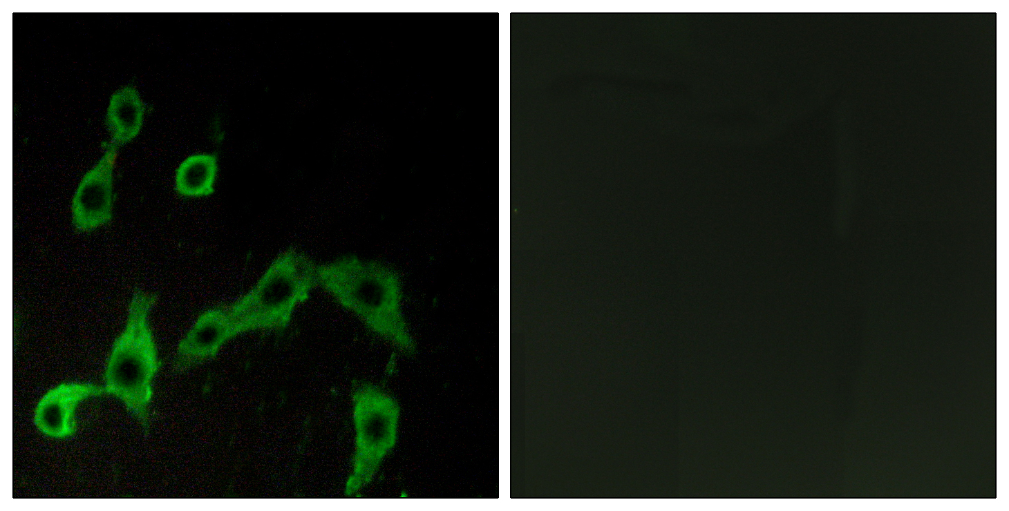
Anti-ADRA2A AntibodyA100622
ApplicationsImmunoFluorescence, ELISA
ReactivityHuman
- SizePrice

Product group Antibodies
Anti-alpha 2a Adrenergic Receptor/ADRA2A Antibody Picoband(r)A00883-3-CARRIER-FREE
ApplicationsFlow Cytometry, Western Blot, ELISA
ReactivityHuman, Mouse, Rat
TargetADRA2A
- SizePrice
Product group Antibodies
Anti-ADRA2A Antibody144-02809
ApplicationsWestern Blot
ReactivityHuman, Mouse
TargetADRA2A
- SizePrice
Product group Antibodies

ADRA2A AntibodyLS-C748957
ApplicationsWestern Blot
ReactivityHuman, Mouse, Rat
TargetADRA2A
- SizePrice
Product group Antibodies
References
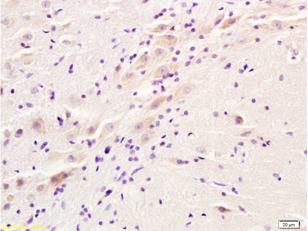
ADRA2 Polyclonal AntibodyBS-1062R
ApplicationsImmunoFluorescence, Western Blot, ELISA, ImmunoCytoChemistry, ImmunoHistoChemistry, ImmunoHistoChemistry Frozen, ImmunoHistoChemistry Paraffin
TargetADRA2A
- SizePrice
Product group Antibodies
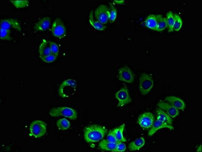
ADRA2A AntibodyCSB-PA001388LA01HU
ApplicationsImmunoFluorescence, ELISA
ReactivityHuman
TargetADRA2A
- SizePrice
Product group Antibodies

Goat anti-ADRA2AEB07121
ApplicationsFlow Cytometry, ImmunoFluorescence, ELISA
ReactivityCanine, Human, Mouse, Rat
TargetADRA2A
- SizePrice
![Boiled and unboiled LNCap whole cell and membrane extracts (30 μg) were separated by 10% SDS-PAGE, and the membrane was blotted with alpha 2a Adrenergic Receptor antibody [HL3723] (GTX641913) diluted at 1:1000. The HRP-conjugated anti-rabbit IgG antibody (GTX213110-01) was used to detect the primary antibody. (WCE: whole cell extract; ME: membrane extract)](https://www.genetex.com/upload/website/prouct_img/normal/GTX641913/GTX641913_T-45663_20250124_WB_Fraction_ub_25020422_270.webp)
Product group Antibodies
ApplicationsWestern Blot
ReactivityHuman, Mouse
TargetADRA2A
- SizePrice